Manganese in PDB 1tbl: H141N Mutant of Rat Liver Arginase I
Enzymatic activity of H141N Mutant of Rat Liver Arginase I
All present enzymatic activity of H141N Mutant of Rat Liver Arginase I:
3.5.3.1;
Protein crystallography data
The structure of H141N Mutant of Rat Liver Arginase I, PDB code: 1tbl
was solved by
E.Cama,
J.D.Cox,
D.E.Ash,
D.W.Christianson,
with X-Ray Crystallography technique. A brief refinement statistics is given in the table below:
Resolution Low / High (Å)
|
45.56 /
3.10
|
Space group
|
P 32
|
Cell size a, b, c (Å), α, β, γ (°)
|
88.332,
88.332,
113.417,
90.00,
90.00,
120.00
|
R / Rfree (%)
|
29.8 /
30.6
|
Manganese Binding Sites:
The binding sites of Manganese atom in the H141N Mutant of Rat Liver Arginase I
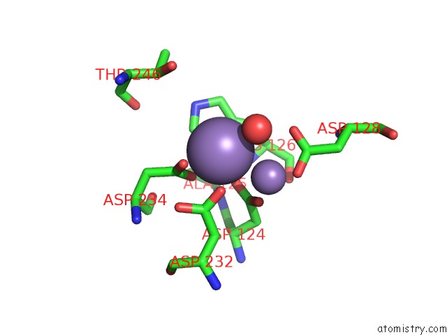
(pdb code 1tbl). This binding sites where shown within
5.0 Angstroms radius around Manganese atom.
In total 6 binding sites of Manganese where determined in the
H141N Mutant of Rat Liver Arginase I, PDB code: 1tbl:
Jump to Manganese binding site number:
1;
2;
3;
4;
5;
6;
Manganese binding site 1 out
of 6 in 1tbl
Go back to
Manganese Binding Sites List in 1tbl
Manganese binding site 1 out
of 6 in the H141N Mutant of Rat Liver Arginase I
Mono view
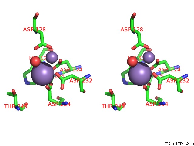
Stereo pair view
|
A full contact list of Manganese with other atoms in the Mn binding
site number 1 of H141N Mutant of Rat Liver Arginase I within 5.0Å range:
probe
|
atom
|
residue
|
distance (Å)
|
B
|
Occ
|
A:Mn1
b:40.3
occ:1.00
|
OD2
|
A:ASP234
|
2.1
|
58.5
|
1.0
|
ND1
|
A:HIS126
|
2.4
|
59.7
|
1.0
|
OD1
|
A:ASP234
|
2.4
|
58.4
|
1.0
|
OD2
|
A:ASP232
|
2.5
|
38.8
|
1.0
|
OD1
|
A:ASP124
|
2.5
|
63.3
|
1.0
|
CG
|
A:ASP234
|
2.5
|
56.9
|
1.0
|
O
|
A:HOH800
|
2.6
|
91.5
|
1.0
|
CE1
|
A:HIS126
|
3.0
|
57.2
|
1.0
|
CG
|
A:ASP124
|
3.2
|
65.3
|
1.0
|
OD2
|
A:ASP124
|
3.3
|
67.5
|
1.0
|
CG
|
A:ASP232
|
3.3
|
40.3
|
1.0
|
CG
|
A:HIS126
|
3.3
|
60.5
|
1.0
|
MN
|
A:MN2
|
3.3
|
59.4
|
1.0
|
OD1
|
A:ASP232
|
3.7
|
38.6
|
1.0
|
CB
|
A:HIS126
|
3.8
|
63.5
|
1.0
|
OD1
|
A:ASP128
|
3.9
|
72.0
|
1.0
|
NE2
|
A:HIS126
|
3.9
|
56.4
|
1.0
|
CB
|
A:ASP234
|
4.0
|
53.0
|
1.0
|
OD2
|
A:ASP128
|
4.1
|
71.9
|
1.0
|
CD2
|
A:HIS126
|
4.1
|
57.6
|
1.0
|
CB
|
A:ASP232
|
4.3
|
43.5
|
1.0
|
CG
|
A:ASP128
|
4.4
|
73.2
|
1.0
|
N
|
A:HIS126
|
4.5
|
69.8
|
1.0
|
CB
|
A:ASP124
|
4.7
|
64.7
|
1.0
|
N
|
A:ALA125
|
4.7
|
64.0
|
1.0
|
CA
|
A:HIS126
|
4.7
|
70.9
|
1.0
|
OG1
|
A:THR246
|
4.9
|
57.4
|
1.0
|
|
Manganese binding site 2 out
of 6 in 1tbl
Go back to
Manganese Binding Sites List in 1tbl
Manganese binding site 2 out
of 6 in the H141N Mutant of Rat Liver Arginase I
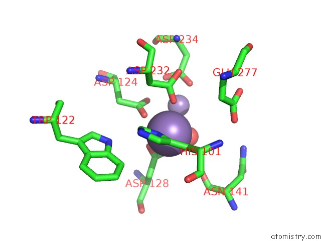
Mono view
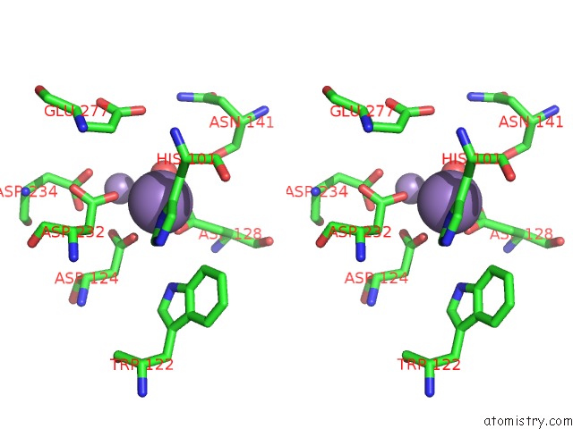
Stereo pair view
|
A full contact list of Manganese with other atoms in the Mn binding
site number 2 of H141N Mutant of Rat Liver Arginase I within 5.0Å range:
probe
|
atom
|
residue
|
distance (Å)
|
B
|
Occ
|
A:Mn2
b:59.4
occ:1.00
|
ND1
|
A:HIS101
|
2.0
|
60.3
|
1.0
|
OD2
|
A:ASP128
|
2.2
|
71.9
|
1.0
|
OD2
|
A:ASP124
|
2.4
|
67.5
|
1.0
|
O
|
A:HOH800
|
2.4
|
91.5
|
1.0
|
OD2
|
A:ASP232
|
2.5
|
38.8
|
1.0
|
CG
|
A:HIS101
|
2.9
|
61.2
|
1.0
|
CG
|
A:ASP128
|
2.9
|
73.2
|
1.0
|
CB
|
A:HIS101
|
3.1
|
62.5
|
1.0
|
CE1
|
A:HIS101
|
3.1
|
58.9
|
1.0
|
OD1
|
A:ASP128
|
3.3
|
72.0
|
1.0
|
MN
|
A:MN1
|
3.3
|
40.3
|
1.0
|
CG
|
A:ASP124
|
3.5
|
65.3
|
1.0
|
CG
|
A:ASP232
|
3.6
|
40.3
|
1.0
|
OD1
|
A:ASP124
|
4.0
|
63.3
|
1.0
|
CB
|
A:ASP128
|
4.1
|
74.4
|
1.0
|
CD2
|
A:HIS101
|
4.1
|
60.4
|
1.0
|
CB
|
A:ASP232
|
4.1
|
43.5
|
1.0
|
NE2
|
A:HIS101
|
4.1
|
60.5
|
1.0
|
O
|
A:ASN141
|
4.2
|
89.4
|
1.0
|
OD2
|
A:ASP234
|
4.4
|
58.5
|
1.0
|
NE1
|
A:TRP122
|
4.5
|
64.6
|
1.0
|
CZ2
|
A:TRP122
|
4.6
|
63.3
|
1.0
|
CA
|
A:HIS101
|
4.6
|
68.5
|
1.0
|
OD1
|
A:ASP232
|
4.7
|
38.6
|
1.0
|
CB
|
A:ASP124
|
4.8
|
64.7
|
1.0
|
CE2
|
A:TRP122
|
4.9
|
63.5
|
1.0
|
OE2
|
A:GLU277
|
5.0
|
72.0
|
1.0
|
CG
|
A:GLU277
|
5.0
|
73.0
|
1.0
|
|
Manganese binding site 3 out
of 6 in 1tbl
Go back to
Manganese Binding Sites List in 1tbl
Manganese binding site 3 out
of 6 in the H141N Mutant of Rat Liver Arginase I
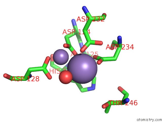
Mono view
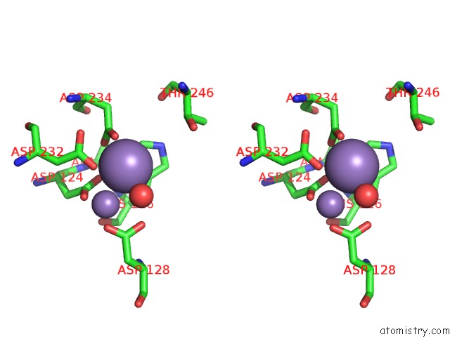
Stereo pair view
|
A full contact list of Manganese with other atoms in the Mn binding
site number 3 of H141N Mutant of Rat Liver Arginase I within 5.0Å range:
probe
|
atom
|
residue
|
distance (Å)
|
B
|
Occ
|
B:Mn3
b:37.0
occ:1.00
|
OD2
|
B:ASP234
|
2.1
|
58.5
|
1.0
|
ND1
|
B:HIS126
|
2.4
|
59.7
|
1.0
|
OD1
|
B:ASP234
|
2.5
|
58.4
|
1.0
|
OD2
|
B:ASP232
|
2.5
|
38.8
|
1.0
|
OD1
|
B:ASP124
|
2.5
|
63.3
|
1.0
|
CG
|
B:ASP234
|
2.6
|
56.9
|
1.0
|
O
|
B:HOH801
|
2.6
|
56.1
|
1.0
|
CE1
|
B:HIS126
|
3.0
|
57.2
|
1.0
|
CG
|
B:ASP124
|
3.3
|
65.3
|
1.0
|
CG
|
B:HIS126
|
3.3
|
60.5
|
1.0
|
OD2
|
B:ASP124
|
3.3
|
67.5
|
1.0
|
CG
|
B:ASP232
|
3.3
|
40.3
|
1.0
|
MN
|
B:MN4
|
3.3
|
56.8
|
1.0
|
OD1
|
B:ASP232
|
3.7
|
38.6
|
1.0
|
CB
|
B:HIS126
|
3.8
|
63.5
|
1.0
|
OD1
|
B:ASP128
|
3.9
|
72.0
|
1.0
|
NE2
|
B:HIS126
|
3.9
|
56.4
|
1.0
|
CB
|
B:ASP234
|
4.0
|
53.0
|
1.0
|
CD2
|
B:HIS126
|
4.1
|
57.6
|
1.0
|
OD2
|
B:ASP128
|
4.1
|
71.9
|
1.0
|
CB
|
B:ASP232
|
4.4
|
43.5
|
1.0
|
CG
|
B:ASP128
|
4.4
|
73.2
|
1.0
|
N
|
B:HIS126
|
4.5
|
69.8
|
1.0
|
CB
|
B:ASP124
|
4.7
|
64.7
|
1.0
|
CA
|
B:HIS126
|
4.8
|
70.9
|
1.0
|
N
|
B:ALA125
|
4.8
|
64.0
|
1.0
|
OG1
|
B:THR246
|
4.9
|
57.4
|
1.0
|
|
Manganese binding site 4 out
of 6 in 1tbl
Go back to
Manganese Binding Sites List in 1tbl
Manganese binding site 4 out
of 6 in the H141N Mutant of Rat Liver Arginase I
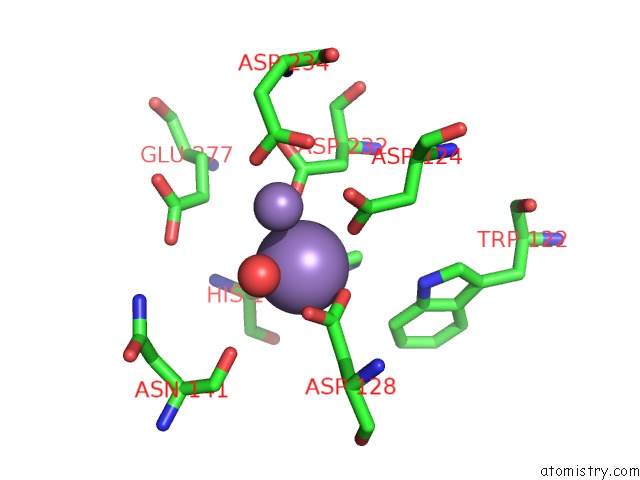
Mono view
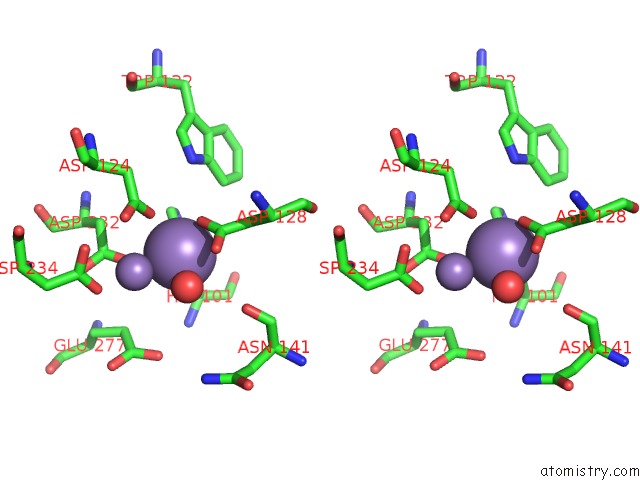
Stereo pair view
|
A full contact list of Manganese with other atoms in the Mn binding
site number 4 of H141N Mutant of Rat Liver Arginase I within 5.0Å range:
probe
|
atom
|
residue
|
distance (Å)
|
B
|
Occ
|
B:Mn4
b:56.8
occ:1.00
|
ND1
|
B:HIS101
|
2.1
|
60.3
|
1.0
|
OD2
|
B:ASP128
|
2.1
|
71.9
|
1.0
|
O
|
B:HOH801
|
2.3
|
56.1
|
1.0
|
OD2
|
B:ASP124
|
2.5
|
67.5
|
1.0
|
OD2
|
B:ASP232
|
2.5
|
38.8
|
1.0
|
CG
|
B:ASP128
|
2.9
|
73.2
|
1.0
|
CG
|
B:HIS101
|
2.9
|
61.2
|
1.0
|
CB
|
B:HIS101
|
3.1
|
62.5
|
1.0
|
CE1
|
B:HIS101
|
3.1
|
58.9
|
1.0
|
OD1
|
B:ASP128
|
3.3
|
72.0
|
1.0
|
MN
|
B:MN3
|
3.3
|
37.0
|
1.0
|
CG
|
B:ASP124
|
3.6
|
65.3
|
1.0
|
CG
|
B:ASP232
|
3.6
|
40.3
|
1.0
|
OD1
|
B:ASP124
|
4.0
|
63.3
|
1.0
|
CB
|
B:ASP128
|
4.0
|
74.4
|
1.0
|
CD2
|
B:HIS101
|
4.1
|
60.4
|
1.0
|
CB
|
B:ASP232
|
4.2
|
43.5
|
1.0
|
NE2
|
B:HIS101
|
4.2
|
60.5
|
1.0
|
O
|
B:ASN141
|
4.2
|
89.4
|
1.0
|
OD2
|
B:ASP234
|
4.4
|
58.5
|
1.0
|
NE1
|
B:TRP122
|
4.6
|
64.6
|
1.0
|
CZ2
|
B:TRP122
|
4.6
|
63.3
|
1.0
|
CA
|
B:HIS101
|
4.6
|
68.5
|
1.0
|
OD1
|
B:ASP232
|
4.7
|
38.6
|
1.0
|
CB
|
B:ASP124
|
4.8
|
64.7
|
1.0
|
OE2
|
B:GLU277
|
5.0
|
72.0
|
1.0
|
CE2
|
B:TRP122
|
5.0
|
63.5
|
1.0
|
CG
|
B:GLU277
|
5.0
|
73.0
|
1.0
|
|
Manganese binding site 5 out
of 6 in 1tbl
Go back to
Manganese Binding Sites List in 1tbl
Manganese binding site 5 out
of 6 in the H141N Mutant of Rat Liver Arginase I
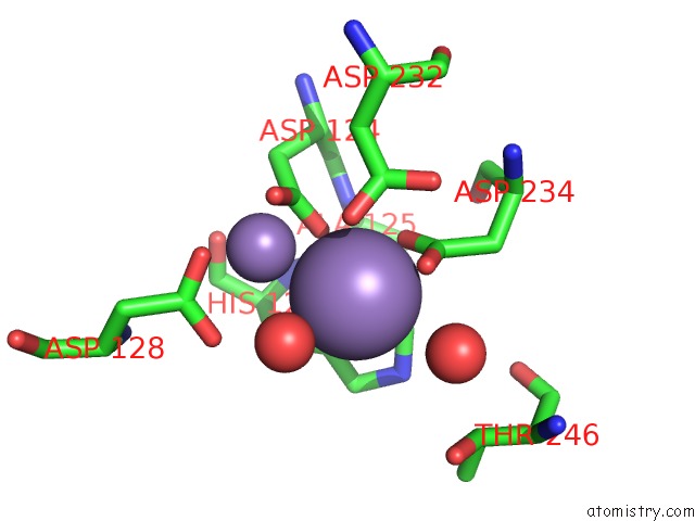
Mono view
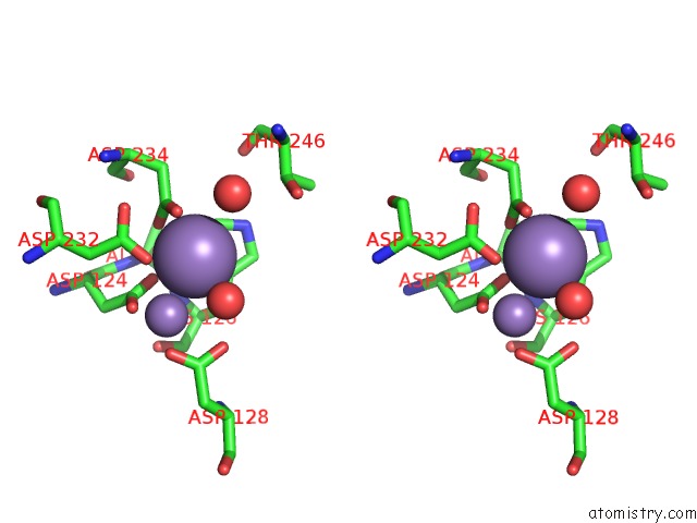
Stereo pair view
|
A full contact list of Manganese with other atoms in the Mn binding
site number 5 of H141N Mutant of Rat Liver Arginase I within 5.0Å range:
probe
|
atom
|
residue
|
distance (Å)
|
B
|
Occ
|
C:Mn5
b:28.4
occ:1.00
|
OD2
|
C:ASP234
|
2.1
|
58.5
|
1.0
|
ND1
|
C:HIS126
|
2.4
|
59.7
|
1.0
|
OD1
|
C:ASP234
|
2.4
|
58.4
|
1.0
|
OD2
|
C:ASP232
|
2.5
|
38.8
|
1.0
|
CG
|
C:ASP234
|
2.5
|
56.9
|
1.0
|
OD1
|
C:ASP124
|
2.5
|
63.3
|
1.0
|
O
|
C:HOH802
|
2.7
|
32.3
|
1.0
|
CE1
|
C:HIS126
|
3.0
|
57.2
|
1.0
|
CG
|
C:HIS126
|
3.3
|
60.5
|
1.0
|
CG
|
C:ASP124
|
3.3
|
65.3
|
1.0
|
CG
|
C:ASP232
|
3.3
|
40.3
|
1.0
|
O
|
C:HOH818
|
3.3
|
21.9
|
1.0
|
OD2
|
C:ASP124
|
3.3
|
67.5
|
1.0
|
MN
|
C:MN320
|
3.3
|
56.5
|
1.0
|
OD1
|
C:ASP232
|
3.7
|
38.6
|
1.0
|
CB
|
C:HIS126
|
3.9
|
63.5
|
1.0
|
NE2
|
C:HIS126
|
3.9
|
56.4
|
1.0
|
OD1
|
C:ASP128
|
3.9
|
72.0
|
1.0
|
CB
|
C:ASP234
|
4.0
|
53.0
|
1.0
|
CD2
|
C:HIS126
|
4.1
|
57.6
|
1.0
|
OD2
|
C:ASP128
|
4.1
|
71.9
|
1.0
|
CB
|
C:ASP232
|
4.4
|
43.5
|
1.0
|
CG
|
C:ASP128
|
4.5
|
73.2
|
1.0
|
N
|
C:HIS126
|
4.5
|
69.8
|
1.0
|
CB
|
C:ASP124
|
4.7
|
64.7
|
1.0
|
N
|
C:ALA125
|
4.8
|
64.0
|
1.0
|
CA
|
C:HIS126
|
4.8
|
70.9
|
1.0
|
OG1
|
C:THR246
|
4.9
|
57.4
|
1.0
|
|
Manganese binding site 6 out
of 6 in 1tbl
Go back to
Manganese Binding Sites List in 1tbl
Manganese binding site 6 out
of 6 in the H141N Mutant of Rat Liver Arginase I
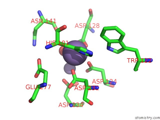
Mono view
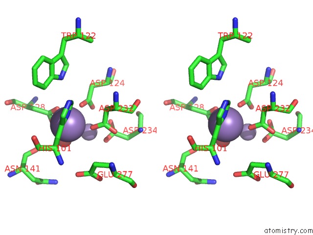
Stereo pair view
|
A full contact list of Manganese with other atoms in the Mn binding
site number 6 of H141N Mutant of Rat Liver Arginase I within 5.0Å range:
probe
|
atom
|
residue
|
distance (Å)
|
B
|
Occ
|
C:Mn320
b:56.5
occ:1.00
|
ND1
|
C:HIS101
|
2.0
|
60.3
|
1.0
|
OD2
|
C:ASP128
|
2.2
|
71.9
|
1.0
|
OD2
|
C:ASP124
|
2.4
|
67.5
|
1.0
|
OD2
|
C:ASP232
|
2.4
|
38.8
|
1.0
|
O
|
C:HOH802
|
2.5
|
32.3
|
1.0
|
CG
|
C:HIS101
|
2.9
|
61.2
|
1.0
|
CG
|
C:ASP128
|
2.9
|
73.2
|
1.0
|
CB
|
C:HIS101
|
3.1
|
62.5
|
1.0
|
CE1
|
C:HIS101
|
3.1
|
58.9
|
1.0
|
OD1
|
C:ASP128
|
3.3
|
72.0
|
1.0
|
MN
|
C:MN5
|
3.3
|
28.4
|
1.0
|
CG
|
C:ASP124
|
3.5
|
65.3
|
1.0
|
CG
|
C:ASP232
|
3.6
|
40.3
|
1.0
|
OD1
|
C:ASP124
|
4.0
|
63.3
|
1.0
|
CD2
|
C:HIS101
|
4.1
|
60.4
|
1.0
|
CB
|
C:ASP128
|
4.1
|
74.4
|
1.0
|
CB
|
C:ASP232
|
4.1
|
43.5
|
1.0
|
NE2
|
C:HIS101
|
4.2
|
60.5
|
1.0
|
O
|
C:ASN141
|
4.3
|
89.4
|
1.0
|
OD2
|
C:ASP234
|
4.4
|
58.5
|
1.0
|
NE1
|
C:TRP122
|
4.6
|
64.6
|
1.0
|
CA
|
C:HIS101
|
4.6
|
68.5
|
1.0
|
OD1
|
C:ASP232
|
4.6
|
38.6
|
1.0
|
CZ2
|
C:TRP122
|
4.6
|
63.3
|
1.0
|
CB
|
C:ASP124
|
4.8
|
64.7
|
1.0
|
CG
|
C:GLU277
|
4.9
|
73.0
|
1.0
|
OE2
|
C:GLU277
|
4.9
|
72.0
|
1.0
|
CE2
|
C:TRP122
|
5.0
|
63.5
|
1.0
|
|
Reference:
D.M.Colleluori,
R.S.Reczkowski,
F.A.Emig,
E.Cama,
J.D.Cox,
L.R.Scolnick,
K.Compher,
K.Jude,
S.Han,
R.E.Viola,
D.W.Christianson,
D.E.Ash.
Probing the Role of the Hyper-Reactive Histidine Residue of Arginase. Arch.Biochem.Biophys. V. 444 15 2005.
ISSN: ISSN 0003-9861
PubMed: 16266687
DOI: 10.1016/J.ABB.2005.09.009
Page generated: Sat Oct 5 12:33:18 2024
|