Manganese »
PDB 1r2m-1sfy »
1r9c »
Manganese in PDB 1r9c: Crystal Structure of Fosfomycin Resistance Protein Fosx From Mesorhizobium Loti
Enzymatic activity of Crystal Structure of Fosfomycin Resistance Protein Fosx From Mesorhizobium Loti
All present enzymatic activity of Crystal Structure of Fosfomycin Resistance Protein Fosx From Mesorhizobium Loti:
2.5.1.1;
2.5.1.1;
Protein crystallography data
The structure of Crystal Structure of Fosfomycin Resistance Protein Fosx From Mesorhizobium Loti, PDB code: 1r9c
was solved by
K.L.Fillgrove,
S.Pakhomova,
M.E.Newcomer,
R.N.Armstrong,
with X-Ray Crystallography technique. A brief refinement statistics is given in the table below:
| Resolution Low / High (Å) | 30.00 / 1.83 |
| Space group | P 21 21 2 |
| Cell size a, b, c (Å), α, β, γ (°) | 45.034, 84.024, 66.861, 90.00, 90.00, 90.00 |
| R / Rfree (%) | 20.2 / 24.3 |
Manganese Binding Sites:
The binding sites of Manganese atom in the Crystal Structure of Fosfomycin Resistance Protein Fosx From Mesorhizobium Loti
(pdb code 1r9c). This binding sites where shown within
5.0 Angstroms radius around Manganese atom.
In total 2 binding sites of Manganese where determined in the Crystal Structure of Fosfomycin Resistance Protein Fosx From Mesorhizobium Loti, PDB code: 1r9c:
Jump to Manganese binding site number: 1; 2;
In total 2 binding sites of Manganese where determined in the Crystal Structure of Fosfomycin Resistance Protein Fosx From Mesorhizobium Loti, PDB code: 1r9c:
Jump to Manganese binding site number: 1; 2;
Manganese binding site 1 out of 2 in 1r9c
Go back to
Manganese binding site 1 out
of 2 in the Crystal Structure of Fosfomycin Resistance Protein Fosx From Mesorhizobium Loti
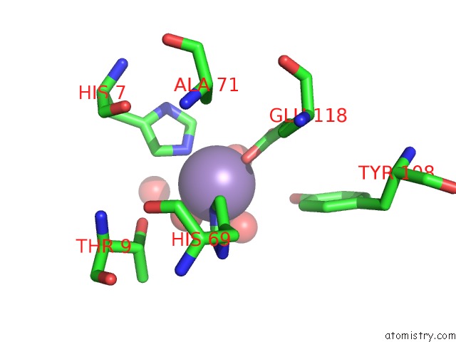
Mono view
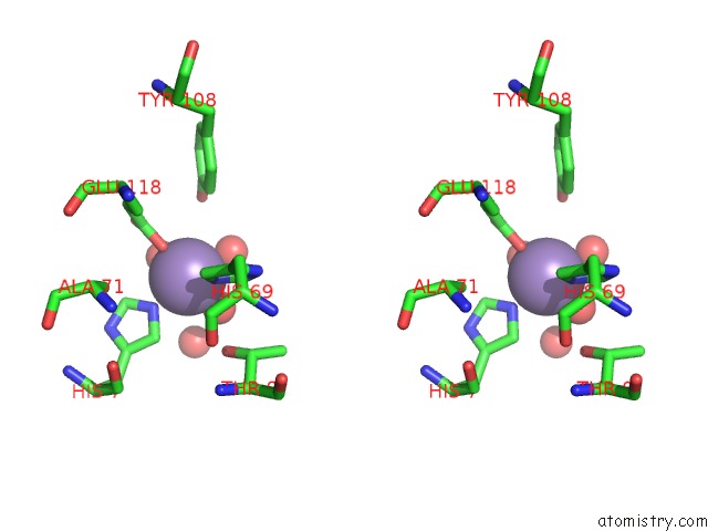
Stereo pair view
Mono view
Stereo pair view
A full contact list of Manganese with other atoms in the Mn binding
site number 1 of Crystal Structure of Fosfomycin Resistance Protein Fosx From Mesorhizobium Loti within 5.0Å range:
|
Manganese binding site 2 out of 2 in 1r9c
Go back to
Manganese binding site 2 out
of 2 in the Crystal Structure of Fosfomycin Resistance Protein Fosx From Mesorhizobium Loti
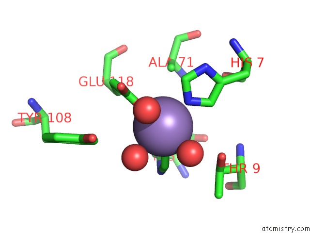
Mono view
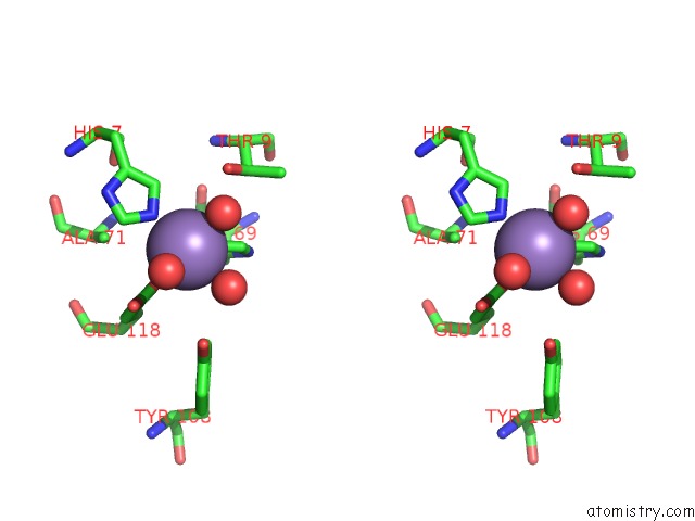
Stereo pair view
Mono view
Stereo pair view
A full contact list of Manganese with other atoms in the Mn binding
site number 2 of Crystal Structure of Fosfomycin Resistance Protein Fosx From Mesorhizobium Loti within 5.0Å range:
|
Reference:
K.L.Fillgrove,
S.Pakhomova,
M.E.Newcomer,
R.N.Armstrong.
Mechanistic Diversity of Fosfomycin Resistance in Pathogenic Microorganisms. J.Am.Chem.Soc. V. 125 15730 2003.
ISSN: ISSN 0002-7863
PubMed: 14677948
DOI: 10.1021/JA039307Z
Page generated: Sat Oct 5 12:19:45 2024
ISSN: ISSN 0002-7863
PubMed: 14677948
DOI: 10.1021/JA039307Z
Last articles
Zn in 9JYWZn in 9IR4
Zn in 9IR3
Zn in 9GMX
Zn in 9GMW
Zn in 9JEJ
Zn in 9ERF
Zn in 9ERE
Zn in 9EGV
Zn in 9EGW