Manganese »
PDB 1pj4-1r1o »
1qoo »
Manganese in PDB 1qoo: Lectin Uea-II Complexed with Nag
Protein crystallography data
The structure of Lectin Uea-II Complexed with Nag, PDB code: 1qoo
was solved by
R.Loris,
H.De Greve,
M.-H.Dao-Thi,
J.Messens,
A.Imberty,
L.Wyns,
with X-Ray Crystallography technique. A brief refinement statistics is given in the table below:
| Resolution Low / High (Å) | 20.00 / 2.75 |
| Space group | P 32 2 1 |
| Cell size a, b, c (Å), α, β, γ (°) | 104.480, 104.480, 175.350, 90.00, 90.00, 120.00 |
| R / Rfree (%) | 17.4 / 20.8 |
Other elements in 1qoo:
The structure of Lectin Uea-II Complexed with Nag also contains other interesting chemical elements:
| Calcium | (Ca) | 4 atoms |
Manganese Binding Sites:
The binding sites of Manganese atom in the Lectin Uea-II Complexed with Nag
(pdb code 1qoo). This binding sites where shown within
5.0 Angstroms radius around Manganese atom.
In total 4 binding sites of Manganese where determined in the Lectin Uea-II Complexed with Nag, PDB code: 1qoo:
Jump to Manganese binding site number: 1; 2; 3; 4;
In total 4 binding sites of Manganese where determined in the Lectin Uea-II Complexed with Nag, PDB code: 1qoo:
Jump to Manganese binding site number: 1; 2; 3; 4;
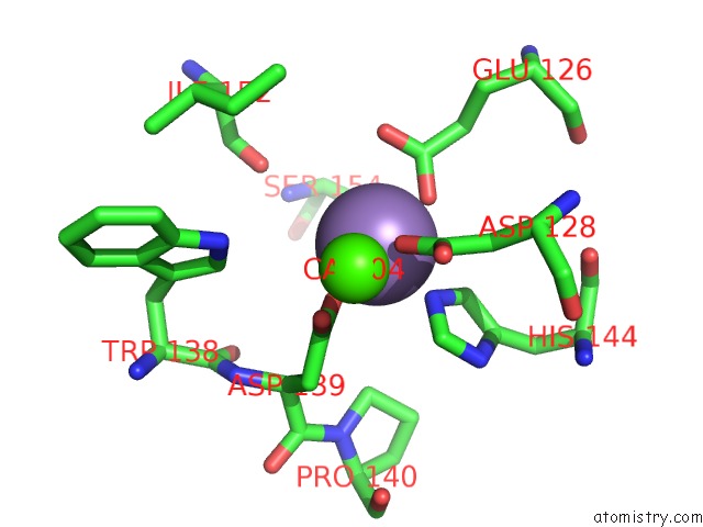
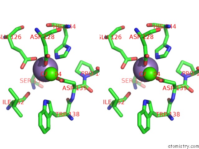
Manganese binding site 1 out of 4 in 1qoo
Go back to
Manganese binding site 1 out
of 4 in the Lectin Uea-II Complexed with Nag
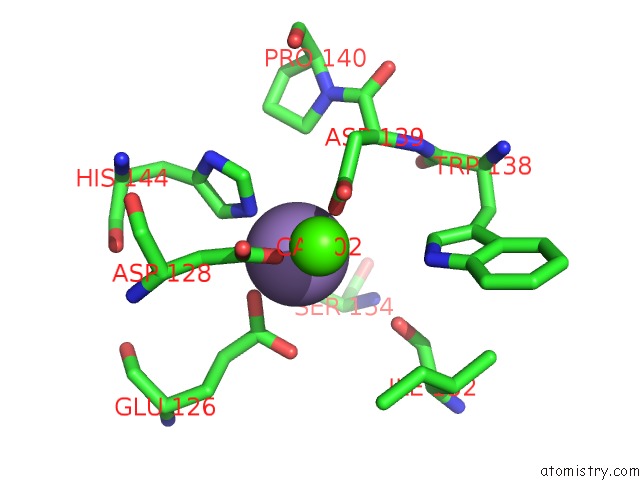
Mono view

Stereo pair view
Mono view
Stereo pair view
A full contact list of Manganese with other atoms in the Mn binding
site number 1 of Lectin Uea-II Complexed with Nag within 5.0Å range:
|
Manganese binding site 2 out of 4 in 1qoo
Go back to
Manganese binding site 2 out
of 4 in the Lectin Uea-II Complexed with Nag
Mono view
Stereo pair view
Mono view
Stereo pair view
A full contact list of Manganese with other atoms in the Mn binding
site number 2 of Lectin Uea-II Complexed with Nag within 5.0Å range:
|
Manganese binding site 3 out of 4 in 1qoo
Go back to
Manganese binding site 3 out
of 4 in the Lectin Uea-II Complexed with Nag

Mono view

Stereo pair view
Mono view
Stereo pair view
A full contact list of Manganese with other atoms in the Mn binding
site number 3 of Lectin Uea-II Complexed with Nag within 5.0Å range:
|
Manganese binding site 4 out of 4 in 1qoo
Go back to
Manganese binding site 4 out
of 4 in the Lectin Uea-II Complexed with Nag

Mono view

Stereo pair view
Mono view
Stereo pair view
A full contact list of Manganese with other atoms in the Mn binding
site number 4 of Lectin Uea-II Complexed with Nag within 5.0Å range:
|
Reference:
R.Loris,
H.De Greve,
M.-H.Dao-Thi,
J.Messens,
A.Imberty,
L.Wyns.
Structural Basis of Carbohydrate Recognition By Lectin II From Ulex Europaeus, A Protein with A Promiscuous Carbohydrate Binding Site J.Mol.Biol. V. 301 987 2000.
ISSN: ISSN 0022-2836
PubMed: 10966800
DOI: 10.1006/JMBI.2000.4016
Page generated: Sat Oct 5 12:14:15 2024
ISSN: ISSN 0022-2836
PubMed: 10966800
DOI: 10.1006/JMBI.2000.4016
Last articles
Zn in 9MJ5Zn in 9HNW
Zn in 9G0L
Zn in 9FNE
Zn in 9DZN
Zn in 9E0I
Zn in 9D32
Zn in 9DAK
Zn in 8ZXC
Zn in 8ZUF