Manganese »
PDB 1pj4-1r1o »
1ql6 »
Manganese in PDB 1ql6: The Catalytic Mechanism of Phosphorylase Kinase Probed By Mutational Studies
Enzymatic activity of The Catalytic Mechanism of Phosphorylase Kinase Probed By Mutational Studies
All present enzymatic activity of The Catalytic Mechanism of Phosphorylase Kinase Probed By Mutational Studies:
2.7.1.38;
2.7.1.38;
Protein crystallography data
The structure of The Catalytic Mechanism of Phosphorylase Kinase Probed By Mutational Studies, PDB code: 1ql6
was solved by
V.T.Skamnaki,
D.J.Owen,
M.E.M.Noble,
E.D.Lowe,
N.G.Oikonomakos,
L.N.Johnson,
with X-Ray Crystallography technique. A brief refinement statistics is given in the table below:
| Resolution Low / High (Å) | 20.00 / 2.40 |
| Space group | P 21 21 21 |
| Cell size a, b, c (Å), α, β, γ (°) | 47.560, 68.171, 112.475, 90.00, 90.00, 90.00 |
| R / Rfree (%) | 24 / 33 |
Manganese Binding Sites:
The binding sites of Manganese atom in the The Catalytic Mechanism of Phosphorylase Kinase Probed By Mutational Studies
(pdb code 1ql6). This binding sites where shown within
5.0 Angstroms radius around Manganese atom.
In total 2 binding sites of Manganese where determined in the The Catalytic Mechanism of Phosphorylase Kinase Probed By Mutational Studies, PDB code: 1ql6:
Jump to Manganese binding site number: 1; 2;
In total 2 binding sites of Manganese where determined in the The Catalytic Mechanism of Phosphorylase Kinase Probed By Mutational Studies, PDB code: 1ql6:
Jump to Manganese binding site number: 1; 2;
Manganese binding site 1 out of 2 in 1ql6
Go back to
Manganese binding site 1 out
of 2 in the The Catalytic Mechanism of Phosphorylase Kinase Probed By Mutational Studies
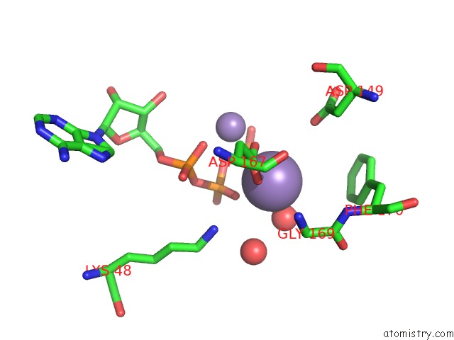
Mono view
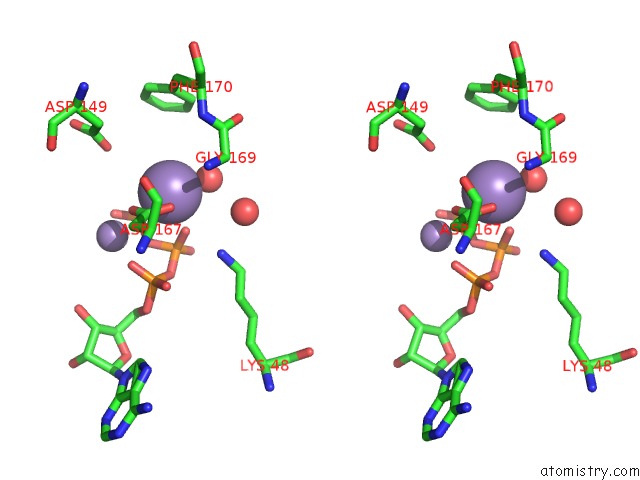
Stereo pair view
Mono view
Stereo pair view
A full contact list of Manganese with other atoms in the Mn binding
site number 1 of The Catalytic Mechanism of Phosphorylase Kinase Probed By Mutational Studies within 5.0Å range:
|
Manganese binding site 2 out of 2 in 1ql6
Go back to
Manganese binding site 2 out
of 2 in the The Catalytic Mechanism of Phosphorylase Kinase Probed By Mutational Studies
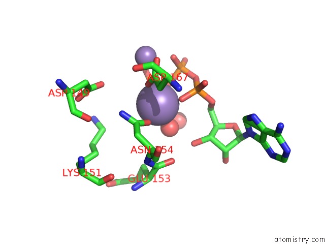
Mono view
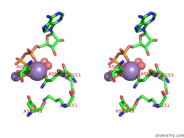
Stereo pair view
Mono view
Stereo pair view
A full contact list of Manganese with other atoms in the Mn binding
site number 2 of The Catalytic Mechanism of Phosphorylase Kinase Probed By Mutational Studies within 5.0Å range:
|
Reference:
V.T.Skamnaki,
D.J.Owen,
M.E.Noble,
E.D.Lowe,
G.Lowe,
N.G.Oikonomakos,
L.N.Johnson.
Catalytic Mechanism of Phosphorylase Kinase Probed By Mutational Studies. Biochemistry V. 38 14718 1999.
ISSN: ISSN 0006-2960
PubMed: 10545198
DOI: 10.1021/BI991454F
Page generated: Sat Oct 5 12:12:54 2024
ISSN: ISSN 0006-2960
PubMed: 10545198
DOI: 10.1021/BI991454F
Last articles
Ca in 2VNVCa in 2VUZ
Ca in 2VUV
Ca in 2VSI
Ca in 2VR0
Ca in 2VS7
Ca in 2VSH
Ca in 2VSF
Ca in 2VRJ
Ca in 2VRG