Manganese »
PDB 1pj4-1r1o »
1pm2 »
Manganese in PDB 1pm2: Crystal Structure of Manganese Substituted R2-D84E (D84E Mutant of the R2 Subunit of E. Coli Ribonucleotide Reductase)
Enzymatic activity of Crystal Structure of Manganese Substituted R2-D84E (D84E Mutant of the R2 Subunit of E. Coli Ribonucleotide Reductase)
All present enzymatic activity of Crystal Structure of Manganese Substituted R2-D84E (D84E Mutant of the R2 Subunit of E. Coli Ribonucleotide Reductase):
1.17.4.1;
1.17.4.1;
Protein crystallography data
The structure of Crystal Structure of Manganese Substituted R2-D84E (D84E Mutant of the R2 Subunit of E. Coli Ribonucleotide Reductase), PDB code: 1pm2
was solved by
W.C.Voegtli,
M.Sommerhalter,
J.Baldwin,
L.Saleh,
J.M.Bollingerjr.,
A.C.Rosenzweig,
with X-Ray Crystallography technique. A brief refinement statistics is given in the table below:
| Resolution Low / High (Å) | 25.00 / 1.80 |
| Space group | P 21 21 21 |
| Cell size a, b, c (Å), α, β, γ (°) | 73.950, 83.940, 114.340, 90.00, 90.00, 90.00 |
| R / Rfree (%) | 18.5 / 21.5 |
Other elements in 1pm2:
The structure of Crystal Structure of Manganese Substituted R2-D84E (D84E Mutant of the R2 Subunit of E. Coli Ribonucleotide Reductase) also contains other interesting chemical elements:
| Mercury | (Hg) | 14 atoms |
Manganese Binding Sites:
The binding sites of Manganese atom in the Crystal Structure of Manganese Substituted R2-D84E (D84E Mutant of the R2 Subunit of E. Coli Ribonucleotide Reductase)
(pdb code 1pm2). This binding sites where shown within
5.0 Angstroms radius around Manganese atom.
In total 4 binding sites of Manganese where determined in the Crystal Structure of Manganese Substituted R2-D84E (D84E Mutant of the R2 Subunit of E. Coli Ribonucleotide Reductase), PDB code: 1pm2:
Jump to Manganese binding site number: 1; 2; 3; 4;
In total 4 binding sites of Manganese where determined in the Crystal Structure of Manganese Substituted R2-D84E (D84E Mutant of the R2 Subunit of E. Coli Ribonucleotide Reductase), PDB code: 1pm2:
Jump to Manganese binding site number: 1; 2; 3; 4;
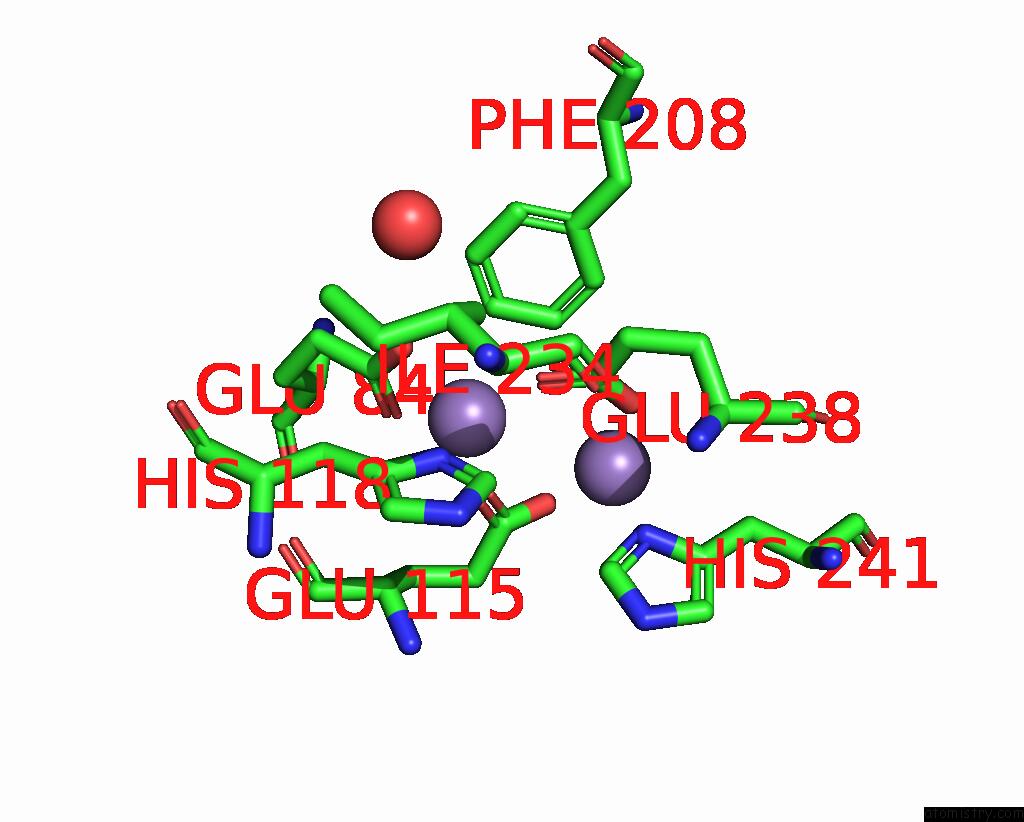
Manganese binding site 1 out of 4 in 1pm2
Go back to
Manganese binding site 1 out
of 4 in the Crystal Structure of Manganese Substituted R2-D84E (D84E Mutant of the R2 Subunit of E. Coli Ribonucleotide Reductase)
Mono view

Stereo pair view
Mono view
Stereo pair view
A full contact list of Manganese with other atoms in the Mn binding
site number 1 of Crystal Structure of Manganese Substituted R2-D84E (D84E Mutant of the R2 Subunit of E. Coli Ribonucleotide Reductase) within 5.0Å range:
|
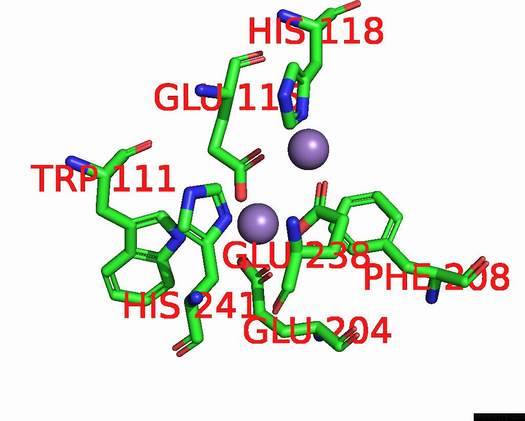
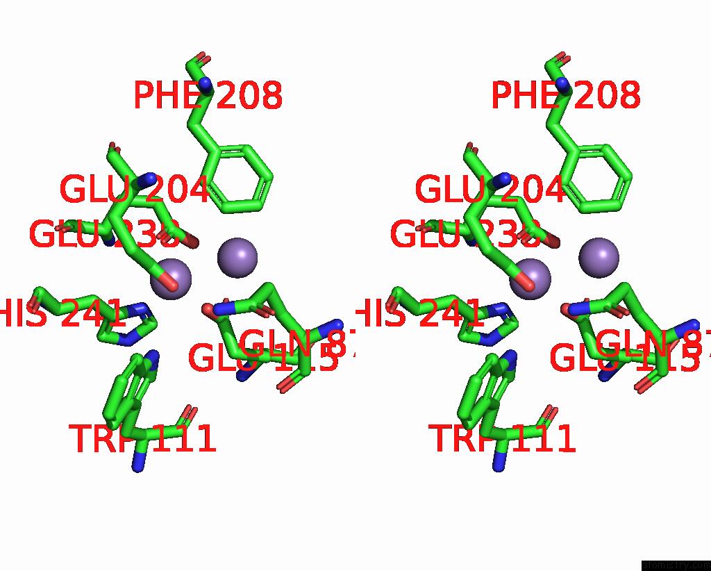
Manganese binding site 2 out of 4 in 1pm2
Go back to
Manganese binding site 2 out
of 4 in the Crystal Structure of Manganese Substituted R2-D84E (D84E Mutant of the R2 Subunit of E. Coli Ribonucleotide Reductase)
Mono view

Stereo pair view
Mono view
Stereo pair view
A full contact list of Manganese with other atoms in the Mn binding
site number 2 of Crystal Structure of Manganese Substituted R2-D84E (D84E Mutant of the R2 Subunit of E. Coli Ribonucleotide Reductase) within 5.0Å range:
|
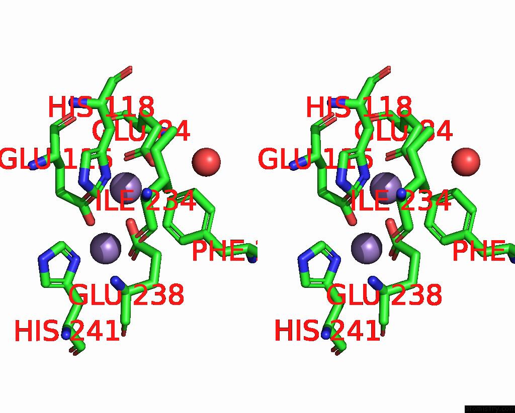
Manganese binding site 3 out of 4 in 1pm2
Go back to
Manganese binding site 3 out
of 4 in the Crystal Structure of Manganese Substituted R2-D84E (D84E Mutant of the R2 Subunit of E. Coli Ribonucleotide Reductase)

Mono view
Stereo pair view
Mono view
Stereo pair view
A full contact list of Manganese with other atoms in the Mn binding
site number 3 of Crystal Structure of Manganese Substituted R2-D84E (D84E Mutant of the R2 Subunit of E. Coli Ribonucleotide Reductase) within 5.0Å range:
|
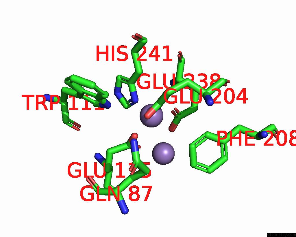
Manganese binding site 4 out of 4 in 1pm2
Go back to
Manganese binding site 4 out
of 4 in the Crystal Structure of Manganese Substituted R2-D84E (D84E Mutant of the R2 Subunit of E. Coli Ribonucleotide Reductase)
Mono view
Stereo pair view
Mono view
Stereo pair view
A full contact list of Manganese with other atoms in the Mn binding
site number 4 of Crystal Structure of Manganese Substituted R2-D84E (D84E Mutant of the R2 Subunit of E. Coli Ribonucleotide Reductase) within 5.0Å range:
|
Reference:
W.C.Voegtli,
M.Sommerhalter,
L.Saleh,
J.Baldwin,
J.M.Bollinger Jr.,
A.C.Rosenzweig.
Variable Coordination Geometries at the Diiron(II) Active Site of Ribonucleotide Reductase R2. J.Am.Chem.Soc. V. 125 15822 2003.
ISSN: ISSN 0002-7863
PubMed: 14677973
DOI: 10.1021/JA0370387
Page generated: Sat Oct 5 12:07:35 2024
ISSN: ISSN 0002-7863
PubMed: 14677973
DOI: 10.1021/JA0370387
Last articles
Zn in 9J0NZn in 9J0O
Zn in 9J0P
Zn in 9FJX
Zn in 9EKB
Zn in 9C0F
Zn in 9CAH
Zn in 9CH0
Zn in 9CH3
Zn in 9CH1