Manganese »
PDB 1o9i-1pj3 »
1phk »
Manganese in PDB 1phk: Two Structures of the Catalytic Domain of Phosphorylase, Kinase: An Active Protein Kinase Complexed with Nucleotide, Substrate-Analogue and Product
Enzymatic activity of Two Structures of the Catalytic Domain of Phosphorylase, Kinase: An Active Protein Kinase Complexed with Nucleotide, Substrate-Analogue and Product
All present enzymatic activity of Two Structures of the Catalytic Domain of Phosphorylase, Kinase: An Active Protein Kinase Complexed with Nucleotide, Substrate-Analogue and Product:
2.7.1.38;
2.7.1.38;
Protein crystallography data
The structure of Two Structures of the Catalytic Domain of Phosphorylase, Kinase: An Active Protein Kinase Complexed with Nucleotide, Substrate-Analogue and Product, PDB code: 1phk
was solved by
D.J.Owen,
M.E.M.Noble,
E.F.Garman,
A.C.Papageorgiou,
L.N.Johnson,
with X-Ray Crystallography technique. A brief refinement statistics is given in the table below:
| Resolution Low / High (Å) | 6.00 / 2.20 |
| Space group | P 21 21 21 |
| Cell size a, b, c (Å), α, β, γ (°) | 47.600, 67.400, 110.800, 90.00, 90.00, 90.00 |
| R / Rfree (%) | 21 / 28.8 |
Manganese Binding Sites:
The binding sites of Manganese atom in the Two Structures of the Catalytic Domain of Phosphorylase, Kinase: An Active Protein Kinase Complexed with Nucleotide, Substrate-Analogue and Product
(pdb code 1phk). This binding sites where shown within
5.0 Angstroms radius around Manganese atom.
In total 2 binding sites of Manganese where determined in the Two Structures of the Catalytic Domain of Phosphorylase, Kinase: An Active Protein Kinase Complexed with Nucleotide, Substrate-Analogue and Product, PDB code: 1phk:
Jump to Manganese binding site number: 1; 2;
In total 2 binding sites of Manganese where determined in the Two Structures of the Catalytic Domain of Phosphorylase, Kinase: An Active Protein Kinase Complexed with Nucleotide, Substrate-Analogue and Product, PDB code: 1phk:
Jump to Manganese binding site number: 1; 2;
Manganese binding site 1 out of 2 in 1phk
Go back to
Manganese binding site 1 out
of 2 in the Two Structures of the Catalytic Domain of Phosphorylase, Kinase: An Active Protein Kinase Complexed with Nucleotide, Substrate-Analogue and Product
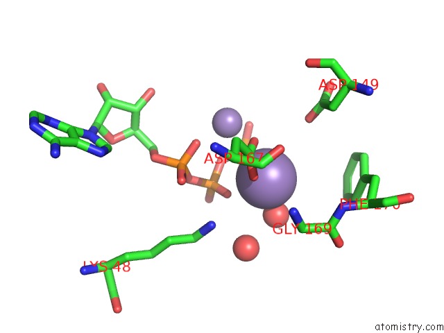
Mono view
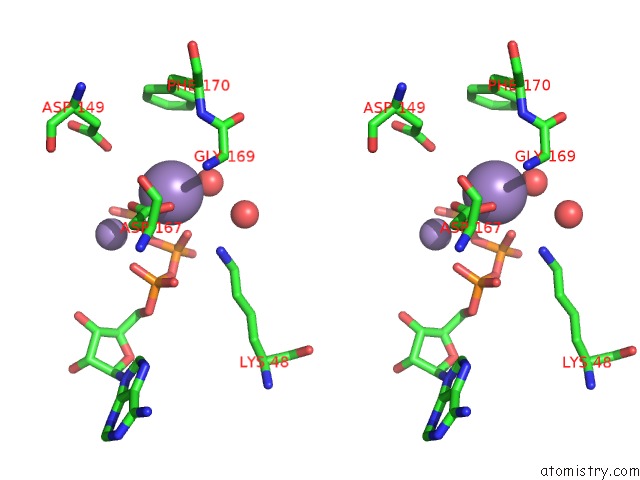
Stereo pair view
Mono view
Stereo pair view
A full contact list of Manganese with other atoms in the Mn binding
site number 1 of Two Structures of the Catalytic Domain of Phosphorylase, Kinase: An Active Protein Kinase Complexed with Nucleotide, Substrate-Analogue and Product within 5.0Å range:
|
Manganese binding site 2 out of 2 in 1phk
Go back to
Manganese binding site 2 out
of 2 in the Two Structures of the Catalytic Domain of Phosphorylase, Kinase: An Active Protein Kinase Complexed with Nucleotide, Substrate-Analogue and Product
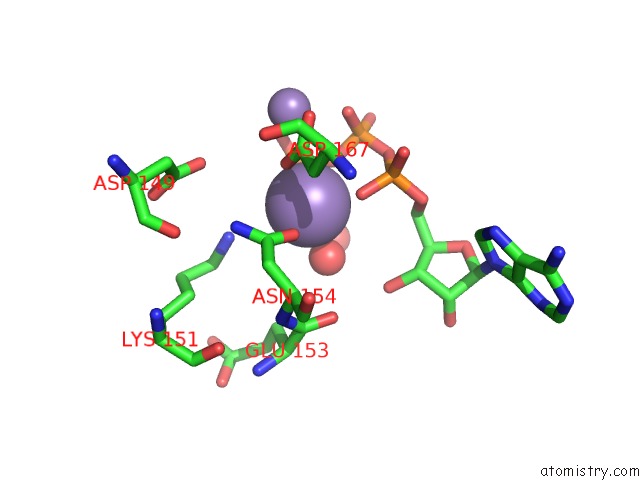
Mono view
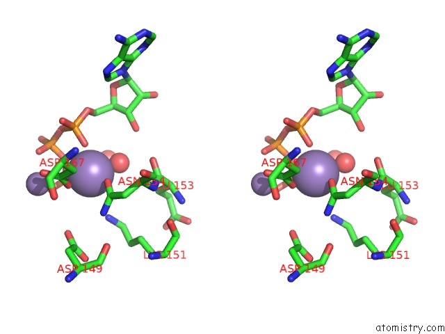
Stereo pair view
Mono view
Stereo pair view
A full contact list of Manganese with other atoms in the Mn binding
site number 2 of Two Structures of the Catalytic Domain of Phosphorylase, Kinase: An Active Protein Kinase Complexed with Nucleotide, Substrate-Analogue and Product within 5.0Å range:
|
Reference:
D.J.Owen,
M.E.Noble,
E.F.Garman,
A.C.Papageorgiou,
L.N.Johnson.
Two Structures of the Catalytic Domain of Phosphorylase Kinase: An Active Protein Kinase Complexed with Substrate Analogue and Product. Structure V. 3 467 1995.
ISSN: ISSN 0969-2126
PubMed: 7663944
DOI: 10.1016/S0969-2126(01)00180-0
Page generated: Sat Oct 5 12:05:16 2024
ISSN: ISSN 0969-2126
PubMed: 7663944
DOI: 10.1016/S0969-2126(01)00180-0
Last articles
Zn in 9J0NZn in 9J0O
Zn in 9J0P
Zn in 9FJX
Zn in 9EKB
Zn in 9C0F
Zn in 9CAH
Zn in 9CH0
Zn in 9CH3
Zn in 9CH1