Manganese »
PDB 1n0n-1o99 »
1nvk »
Manganese in PDB 1nvk: T4 Phage Bgt in Complex with Udp and A MN2+ Ion at 1.8 A Resolution
Enzymatic activity of T4 Phage Bgt in Complex with Udp and A MN2+ Ion at 1.8 A Resolution
All present enzymatic activity of T4 Phage Bgt in Complex with Udp and A MN2+ Ion at 1.8 A Resolution:
2.4.1.27;
2.4.1.27;
Protein crystallography data
The structure of T4 Phage Bgt in Complex with Udp and A MN2+ Ion at 1.8 A Resolution, PDB code: 1nvk
was solved by
L.Lariviere,
J.Kurzeck,
V.Gueguen-Chaignon,
W.Rueger,
S.Morera,
with X-Ray Crystallography technique. A brief refinement statistics is given in the table below:
| Resolution Low / High (Å) | 20.00 / 1.80 |
| Space group | P 1 21 1 |
| Cell size a, b, c (Å), α, β, γ (°) | 47.808, 71.400, 62.000, 90.00, 91.65, 90.00 |
| R / Rfree (%) | 17.4 / 21 |
Manganese Binding Sites:
The binding sites of Manganese atom in the T4 Phage Bgt in Complex with Udp and A MN2+ Ion at 1.8 A Resolution
(pdb code 1nvk). This binding sites where shown within
5.0 Angstroms radius around Manganese atom.
In total only one binding site of Manganese was determined in the T4 Phage Bgt in Complex with Udp and A MN2+ Ion at 1.8 A Resolution, PDB code: 1nvk:
In total only one binding site of Manganese was determined in the T4 Phage Bgt in Complex with Udp and A MN2+ Ion at 1.8 A Resolution, PDB code: 1nvk:
Manganese binding site 1 out of 1 in 1nvk
Go back to
Manganese binding site 1 out
of 1 in the T4 Phage Bgt in Complex with Udp and A MN2+ Ion at 1.8 A Resolution
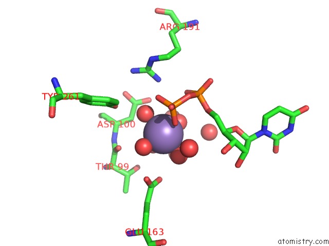
Mono view
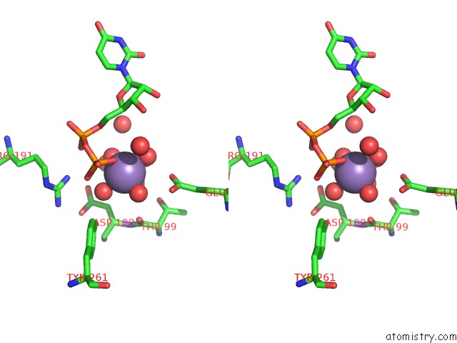
Stereo pair view
Mono view
Stereo pair view
A full contact list of Manganese with other atoms in the Mn binding
site number 1 of T4 Phage Bgt in Complex with Udp and A MN2+ Ion at 1.8 A Resolution within 5.0Å range:
|
Reference:
L.Lariviere,
V.Gueguen-Chaignon,
S.Morera.
Crystal Structures of the T4 Phage Beta-Glucosyltransferase and the D100A Mutant in Complex with Udp-Glucose: Glucose Binding and Identification of the Catalytic Base For A Direct Displacement Mechanism J.Mol.Biol. V. 330 1077 2003.
ISSN: ISSN 0022-2836
PubMed: 12860129
DOI: 10.1016/S0022-2836(03)00635-1
Page generated: Sat Oct 5 11:53:11 2024
ISSN: ISSN 0022-2836
PubMed: 12860129
DOI: 10.1016/S0022-2836(03)00635-1
Last articles
Cl in 5WDJCl in 5WE8
Cl in 5WBG
Cl in 5WDW
Cl in 5WE7
Cl in 5WBV
Cl in 5WDC
Cl in 5WCT
Cl in 5WCI
Cl in 5WBQ