Manganese »
PDB 1n0n-1o99 »
1n51 »
Manganese in PDB 1n51: Aminopeptidase P in Complex with the Inhibitor Apstatin
Enzymatic activity of Aminopeptidase P in Complex with the Inhibitor Apstatin
All present enzymatic activity of Aminopeptidase P in Complex with the Inhibitor Apstatin:
3.4.11.9;
3.4.11.9;
Protein crystallography data
The structure of Aminopeptidase P in Complex with the Inhibitor Apstatin, PDB code: 1n51
was solved by
S.C.Graham,
M.J.Maher,
M.H.Lee,
W.H.Simmons,
H.C.Freeman,
J.M.Guss,
with X-Ray Crystallography technique. A brief refinement statistics is given in the table below:
| Resolution Low / High (Å) | 19.90 / 2.30 |
| Space group | I 41 2 2 |
| Cell size a, b, c (Å), α, β, γ (°) | 139.319, 139.319, 231.005, 90.00, 90.00, 90.00 |
| R / Rfree (%) | 17.9 / 20.4 |
Manganese Binding Sites:
The binding sites of Manganese atom in the Aminopeptidase P in Complex with the Inhibitor Apstatin
(pdb code 1n51). This binding sites where shown within
5.0 Angstroms radius around Manganese atom.
In total 3 binding sites of Manganese where determined in the Aminopeptidase P in Complex with the Inhibitor Apstatin, PDB code: 1n51:
Jump to Manganese binding site number: 1; 2; 3;
In total 3 binding sites of Manganese where determined in the Aminopeptidase P in Complex with the Inhibitor Apstatin, PDB code: 1n51:
Jump to Manganese binding site number: 1; 2; 3;
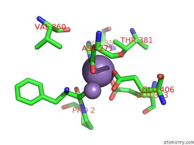
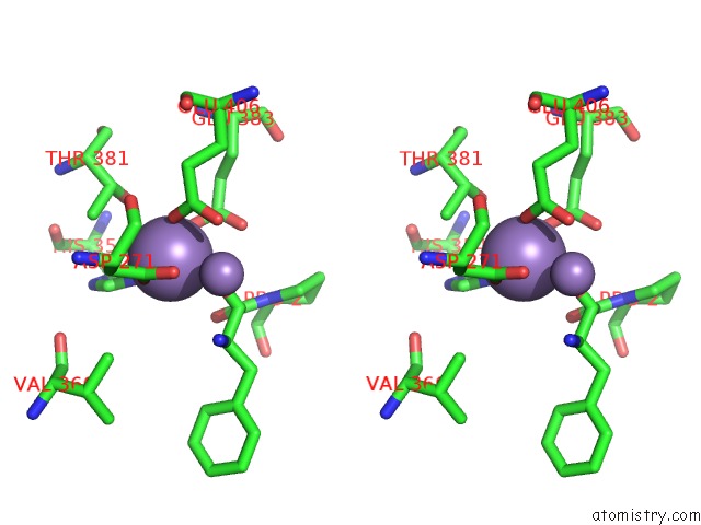
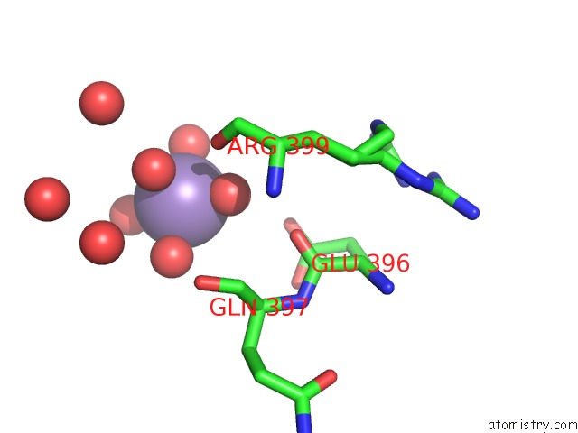
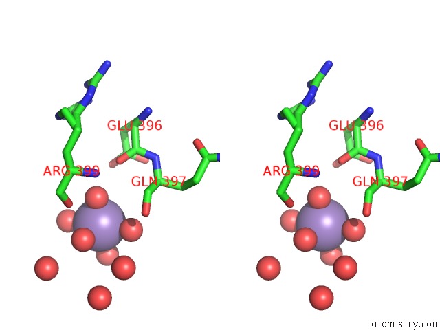
Manganese binding site 1 out of 3 in 1n51
Go back to
Manganese binding site 1 out
of 3 in the Aminopeptidase P in Complex with the Inhibitor Apstatin
Mono view
Stereo pair view
Mono view
Stereo pair view
A full contact list of Manganese with other atoms in the Mn binding
site number 1 of Aminopeptidase P in Complex with the Inhibitor Apstatin within 5.0Å range:
|
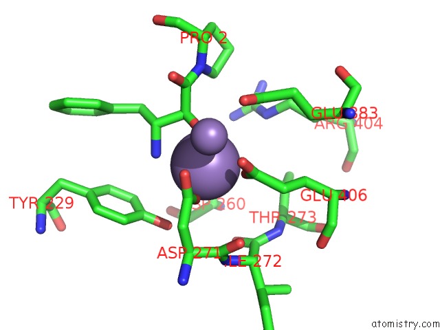
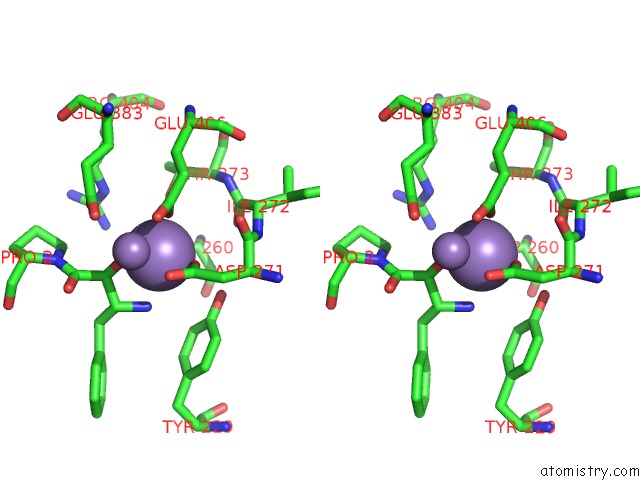
Manganese binding site 2 out of 3 in 1n51
Go back to
Manganese binding site 2 out
of 3 in the Aminopeptidase P in Complex with the Inhibitor Apstatin
Mono view
Stereo pair view
Mono view
Stereo pair view
A full contact list of Manganese with other atoms in the Mn binding
site number 2 of Aminopeptidase P in Complex with the Inhibitor Apstatin within 5.0Å range:
|
Manganese binding site 3 out of 3 in 1n51
Go back to
Manganese binding site 3 out
of 3 in the Aminopeptidase P in Complex with the Inhibitor Apstatin
Mono view
Stereo pair view
Mono view
Stereo pair view
A full contact list of Manganese with other atoms in the Mn binding
site number 3 of Aminopeptidase P in Complex with the Inhibitor Apstatin within 5.0Å range:
|
Reference:
S.C.Graham,
M.J.Maher,
W.H.Simmons,
H.C.Freeman,
J.M.Guss.
Structure of Escherichia Coli Aminopeptidase P in Complex with the Inhibitor Apstatin. Acta Crystallogr.,Sect.D V. 60 1770 2004.
ISSN: ISSN 0907-4449
PubMed: 15388923
DOI: 10.1107/S0907444904018724
Page generated: Sat Oct 5 11:50:08 2024
ISSN: ISSN 0907-4449
PubMed: 15388923
DOI: 10.1107/S0907444904018724
Last articles
Zn in 9MJ5Zn in 9HNW
Zn in 9G0L
Zn in 9FNE
Zn in 9DZN
Zn in 9E0I
Zn in 9D32
Zn in 9DAK
Zn in 8ZXC
Zn in 8ZUF