Manganese »
PDB 1n0n-1o99 »
1n4w »
Manganese in PDB 1n4w: Atomic Resolution Structure of Cholesterol Oxidase @ pH 7.3 (Streptomyces Sp. Sa-Coo)
Enzymatic activity of Atomic Resolution Structure of Cholesterol Oxidase @ pH 7.3 (Streptomyces Sp. Sa-Coo)
All present enzymatic activity of Atomic Resolution Structure of Cholesterol Oxidase @ pH 7.3 (Streptomyces Sp. Sa-Coo):
1.1.3.6;
1.1.3.6;
Protein crystallography data
The structure of Atomic Resolution Structure of Cholesterol Oxidase @ pH 7.3 (Streptomyces Sp. Sa-Coo), PDB code: 1n4w
was solved by
A.Vrielink,
P.I.Lario,
with X-Ray Crystallography technique. A brief refinement statistics is given in the table below:
| Resolution Low / High (Å) | 31.30 / 0.92 |
| Space group | P 1 21 1 |
| Cell size a, b, c (Å), α, β, γ (°) | 51.343, 72.959, 63.045, 90.00, 105.25, 90.00 |
| R / Rfree (%) | 10.3 / 12.2 |
Manganese Binding Sites:
The binding sites of Manganese atom in the Atomic Resolution Structure of Cholesterol Oxidase @ pH 7.3 (Streptomyces Sp. Sa-Coo)
(pdb code 1n4w). This binding sites where shown within
5.0 Angstroms radius around Manganese atom.
In total only one binding site of Manganese was determined in the Atomic Resolution Structure of Cholesterol Oxidase @ pH 7.3 (Streptomyces Sp. Sa-Coo), PDB code: 1n4w:
In total only one binding site of Manganese was determined in the Atomic Resolution Structure of Cholesterol Oxidase @ pH 7.3 (Streptomyces Sp. Sa-Coo), PDB code: 1n4w:
Manganese binding site 1 out of 1 in 1n4w
Go back to
Manganese binding site 1 out
of 1 in the Atomic Resolution Structure of Cholesterol Oxidase @ pH 7.3 (Streptomyces Sp. Sa-Coo)
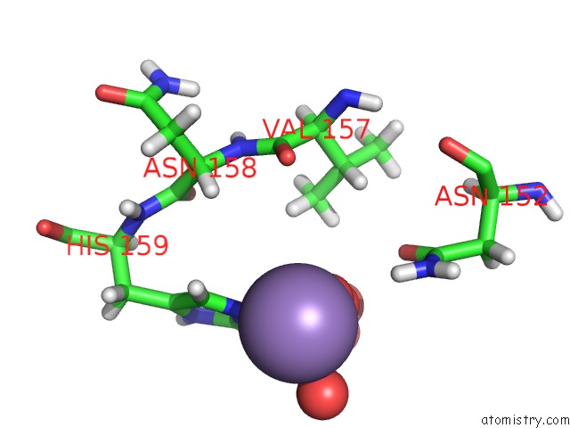
Mono view
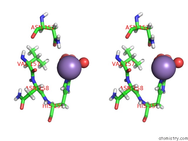
Stereo pair view
Mono view
Stereo pair view
A full contact list of Manganese with other atoms in the Mn binding
site number 1 of Atomic Resolution Structure of Cholesterol Oxidase @ pH 7.3 (Streptomyces Sp. Sa-Coo) within 5.0Å range:
|
Reference:
A.Y.Lyubimov,
P.I.Lario,
I.Moustafa,
A.Vrielink.
Atomic Resolution Crystallography Reveals How Changes in pH Shape the Protein Microenvironment Nat.Chem.Biol. V. 2 259 2006.
ISSN: ISSN 1552-4450
PubMed: 16604066
DOI: 10.1038/NCHEMBIO784
Page generated: Sat Oct 5 11:50:01 2024
ISSN: ISSN 1552-4450
PubMed: 16604066
DOI: 10.1038/NCHEMBIO784
Last articles
Zn in 9J0NZn in 9J0O
Zn in 9J0P
Zn in 9FJX
Zn in 9EKB
Zn in 9C0F
Zn in 9CAH
Zn in 9CH0
Zn in 9CH3
Zn in 9CH1