Manganese »
PDB 1lu1-1n0j »
1mvo »
Manganese in PDB 1mvo: Crystal Structure of the Phop Receiver Domain From Bacillus Subtilis
Protein crystallography data
The structure of Crystal Structure of the Phop Receiver Domain From Bacillus Subtilis, PDB code: 1mvo
was solved by
C.Birck,
Y.Chen,
F.M.Hulett,
J.P.Samama,
Structural Proteomicsin Europe (Spine),
with X-Ray Crystallography technique. A brief refinement statistics is given in the table below:
| Resolution Low / High (Å) | 27.00 / 1.60 |
| Space group | P 41 21 2 |
| Cell size a, b, c (Å), α, β, γ (°) | 45.704, 45.704, 134.811, 90.00, 90.00, 90.00 |
| R / Rfree (%) | 18.6 / 23.2 |
Other elements in 1mvo:
The structure of Crystal Structure of the Phop Receiver Domain From Bacillus Subtilis also contains other interesting chemical elements:
| Sodium | (Na) | 2 atoms |
Manganese Binding Sites:
The binding sites of Manganese atom in the Crystal Structure of the Phop Receiver Domain From Bacillus Subtilis
(pdb code 1mvo). This binding sites where shown within
5.0 Angstroms radius around Manganese atom.
In total only one binding site of Manganese was determined in the Crystal Structure of the Phop Receiver Domain From Bacillus Subtilis, PDB code: 1mvo:
In total only one binding site of Manganese was determined in the Crystal Structure of the Phop Receiver Domain From Bacillus Subtilis, PDB code: 1mvo:
Manganese binding site 1 out of 1 in 1mvo
Go back to
Manganese binding site 1 out
of 1 in the Crystal Structure of the Phop Receiver Domain From Bacillus Subtilis
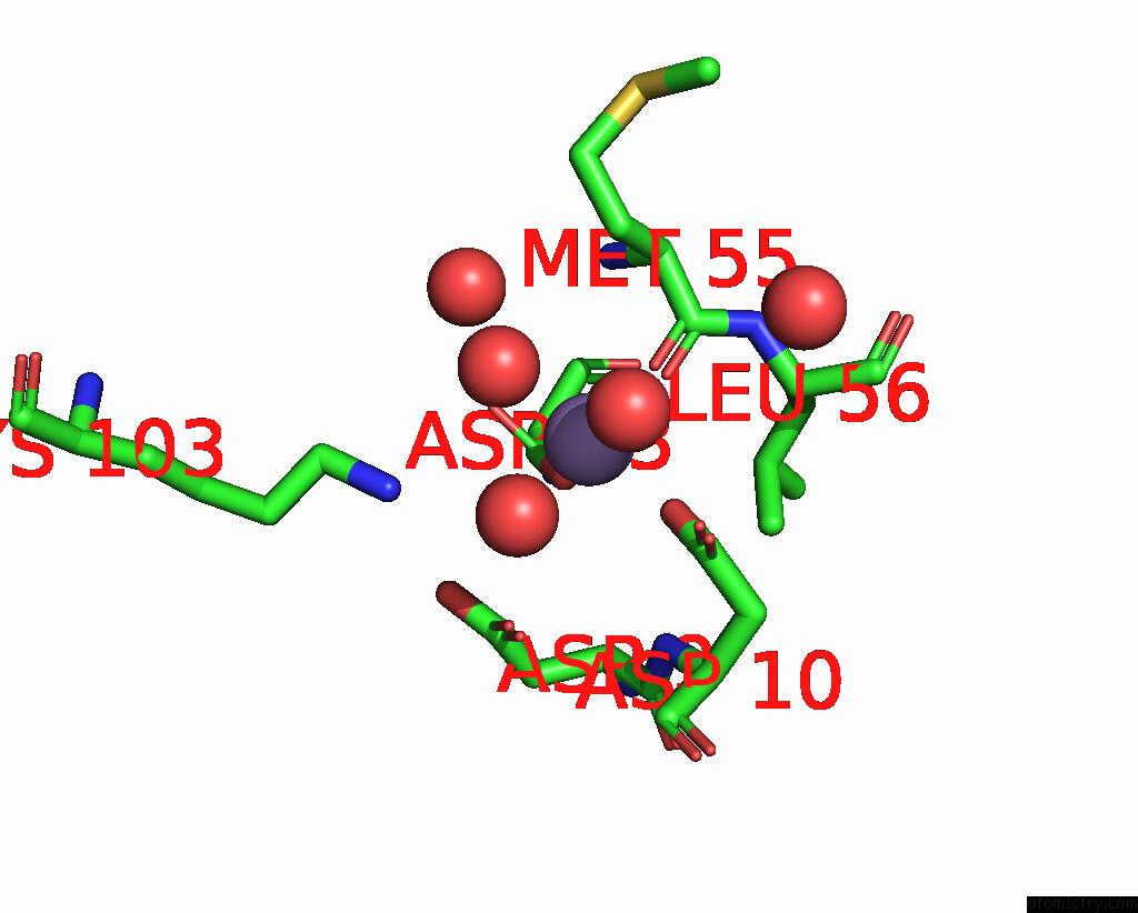
Mono view
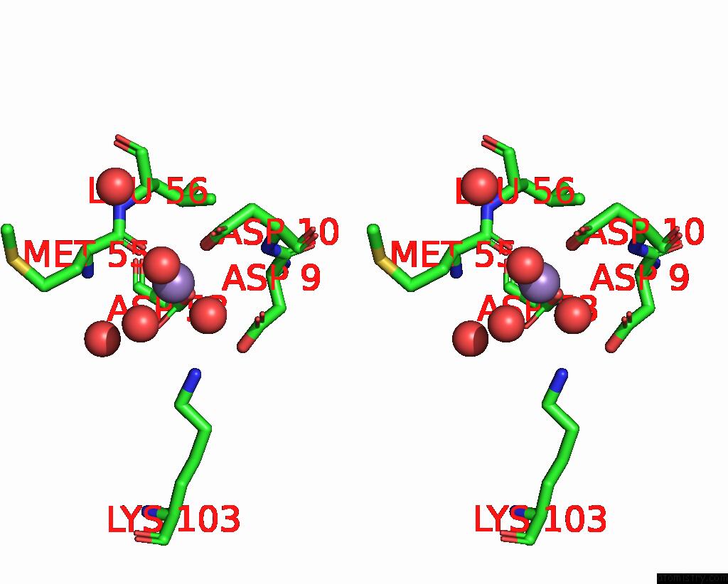
Stereo pair view
Mono view
Stereo pair view
A full contact list of Manganese with other atoms in the Mn binding
site number 1 of Crystal Structure of the Phop Receiver Domain From Bacillus Subtilis within 5.0Å range:
|
Reference:
C.Birck,
Y.Chen,
F.M.Hulett,
J.P.Samama.
The Crystal Structure of the Phosphorylation Domain in Phop Reveals A Functional Tandem Association Mediated By An Asymmetric Interface J.Bacteriol. V. 185 254 2003.
ISSN: ISSN 0021-9193
PubMed: 12486062
DOI: 10.1128/JB.185.1.254-261.2003
Page generated: Sat Oct 5 11:46:04 2024
ISSN: ISSN 0021-9193
PubMed: 12486062
DOI: 10.1128/JB.185.1.254-261.2003
Last articles
Fe in 2YXOFe in 2YRS
Fe in 2YXC
Fe in 2YNM
Fe in 2YVJ
Fe in 2YP1
Fe in 2YU2
Fe in 2YU1
Fe in 2YQB
Fe in 2YOO