Manganese »
PDB 1lu1-1n0j »
1mus »
Manganese in PDB 1mus: Crystal Structure of TN5 Transposase Complexed with Resolved Outside End Dna
Protein crystallography data
The structure of Crystal Structure of TN5 Transposase Complexed with Resolved Outside End Dna, PDB code: 1mus
was solved by
H.M.Holden,
J.B.Thoden,
M.Steiniger-White,
W.S.Reznikoff,
S.Lovell,
I.Rayment,
with X-Ray Crystallography technique. A brief refinement statistics is given in the table below:
| Resolution Low / High (Å) | 30.00 / 1.90 |
| Space group | P 65 2 2 |
| Cell size a, b, c (Å), α, β, γ (°) | 112.700, 112.700, 235.900, 90.00, 90.00, 120.00 |
| R / Rfree (%) | 19 / 24.7 |
Other elements in 1mus:
The structure of Crystal Structure of TN5 Transposase Complexed with Resolved Outside End Dna also contains other interesting chemical elements:
| Magnesium | (Mg) | 2 atoms |
Manganese Binding Sites:
The binding sites of Manganese atom in the Crystal Structure of TN5 Transposase Complexed with Resolved Outside End Dna
(pdb code 1mus). This binding sites where shown within
5.0 Angstroms radius around Manganese atom.
In total 2 binding sites of Manganese where determined in the Crystal Structure of TN5 Transposase Complexed with Resolved Outside End Dna, PDB code: 1mus:
Jump to Manganese binding site number: 1; 2;
In total 2 binding sites of Manganese where determined in the Crystal Structure of TN5 Transposase Complexed with Resolved Outside End Dna, PDB code: 1mus:
Jump to Manganese binding site number: 1; 2;
Manganese binding site 1 out of 2 in 1mus
Go back to
Manganese binding site 1 out
of 2 in the Crystal Structure of TN5 Transposase Complexed with Resolved Outside End Dna
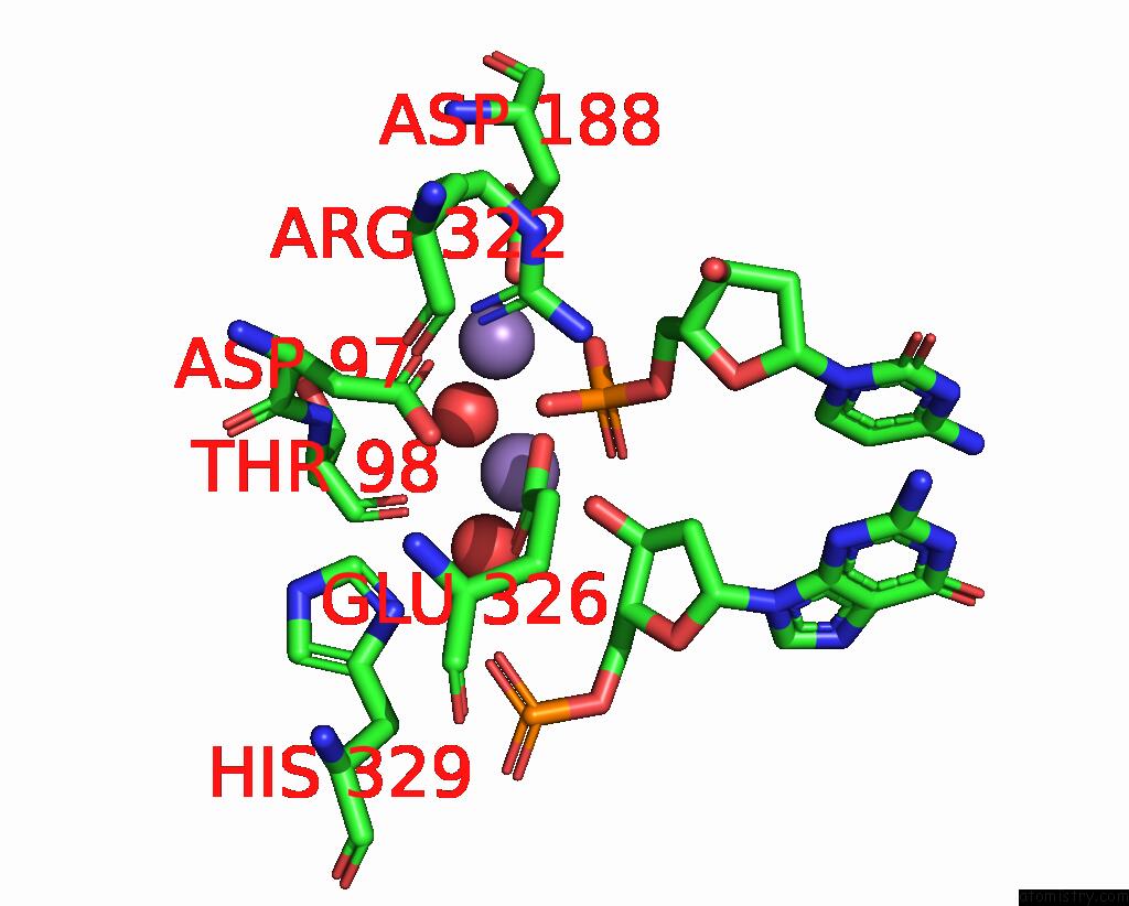
Mono view
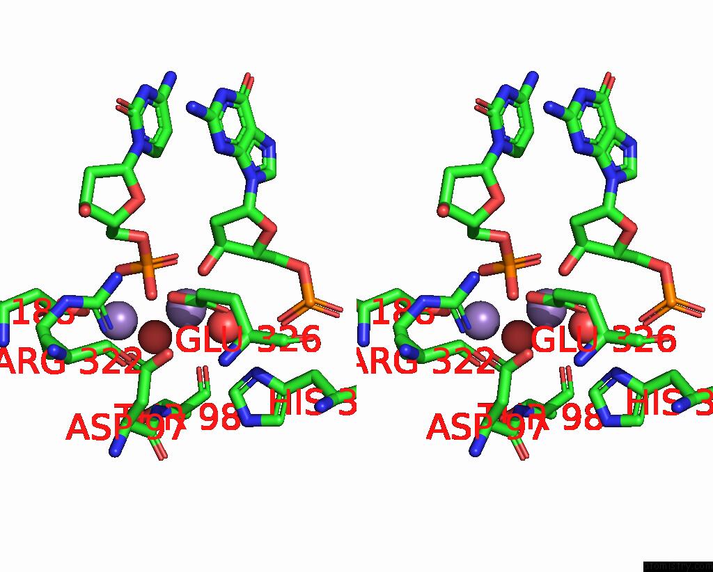
Stereo pair view
Mono view
Stereo pair view
A full contact list of Manganese with other atoms in the Mn binding
site number 1 of Crystal Structure of TN5 Transposase Complexed with Resolved Outside End Dna within 5.0Å range:
|
Manganese binding site 2 out of 2 in 1mus
Go back to
Manganese binding site 2 out
of 2 in the Crystal Structure of TN5 Transposase Complexed with Resolved Outside End Dna
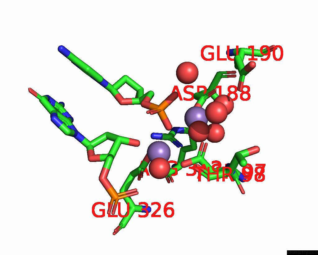
Mono view
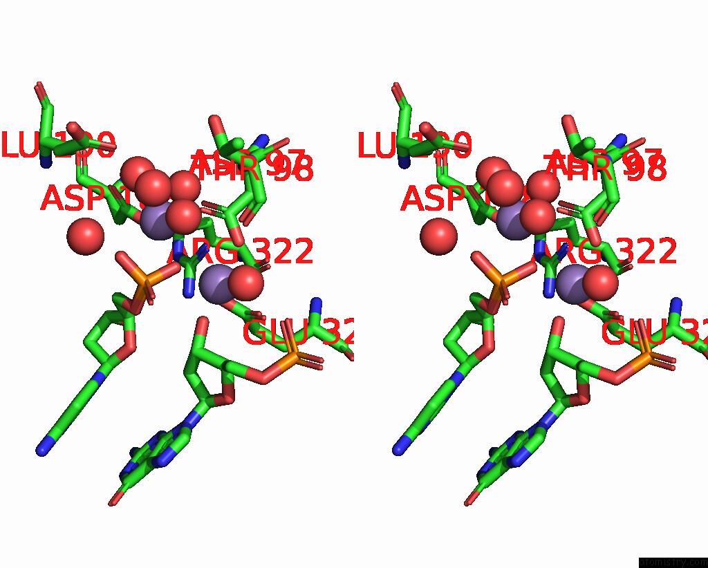
Stereo pair view
Mono view
Stereo pair view
A full contact list of Manganese with other atoms in the Mn binding
site number 2 of Crystal Structure of TN5 Transposase Complexed with Resolved Outside End Dna within 5.0Å range:
|
Reference:
M.Steiniger-White,
I.Rayment,
W.S.Reznikoff.
Structure/Function Insights Into TN5 Transposition. Curr.Opin.Struct.Biol. V. 14 50 2004.
ISSN: ISSN 0959-440X
PubMed: 15102449
DOI: 10.1016/J.SBI.2004.01.008
Page generated: Sat Oct 5 11:46:01 2024
ISSN: ISSN 0959-440X
PubMed: 15102449
DOI: 10.1016/J.SBI.2004.01.008
Last articles
Zn in 9MJ5Zn in 9HNW
Zn in 9G0L
Zn in 9FNE
Zn in 9DZN
Zn in 9E0I
Zn in 9D32
Zn in 9DAK
Zn in 8ZXC
Zn in 8ZUF