Manganese »
PDB 1lu1-1n0j »
1lzi »
Manganese in PDB 1lzi: Glycosyltransferase A + Udp + H Antigen Acceptor
Enzymatic activity of Glycosyltransferase A + Udp + H Antigen Acceptor
All present enzymatic activity of Glycosyltransferase A + Udp + H Antigen Acceptor:
2.4.1.40;
2.4.1.40;
Protein crystallography data
The structure of Glycosyltransferase A + Udp + H Antigen Acceptor, PDB code: 1lzi
was solved by
S.I.Patenaude,
N.O.L.Seto,
S.N.Borisova,
A.Szpacenko,
S.L.Marcus,
M.M.Palcic,
S.V.Evans,
with X-Ray Crystallography technique. A brief refinement statistics is given in the table below:
| Resolution Low / High (Å) | 20.00 / 1.35 |
| Space group | C 2 2 21 |
| Cell size a, b, c (Å), α, β, γ (°) | 52.810, 149.580, 79.970, 90.00, 90.00, 90.00 |
| R / Rfree (%) | 17.9 / 20 |
Other elements in 1lzi:
The structure of Glycosyltransferase A + Udp + H Antigen Acceptor also contains other interesting chemical elements:
| Mercury | (Hg) | 5 atoms |
Manganese Binding Sites:
The binding sites of Manganese atom in the Glycosyltransferase A + Udp + H Antigen Acceptor
(pdb code 1lzi). This binding sites where shown within
5.0 Angstroms radius around Manganese atom.
In total only one binding site of Manganese was determined in the Glycosyltransferase A + Udp + H Antigen Acceptor, PDB code: 1lzi:
In total only one binding site of Manganese was determined in the Glycosyltransferase A + Udp + H Antigen Acceptor, PDB code: 1lzi:
Manganese binding site 1 out of 1 in 1lzi
Go back to
Manganese binding site 1 out
of 1 in the Glycosyltransferase A + Udp + H Antigen Acceptor
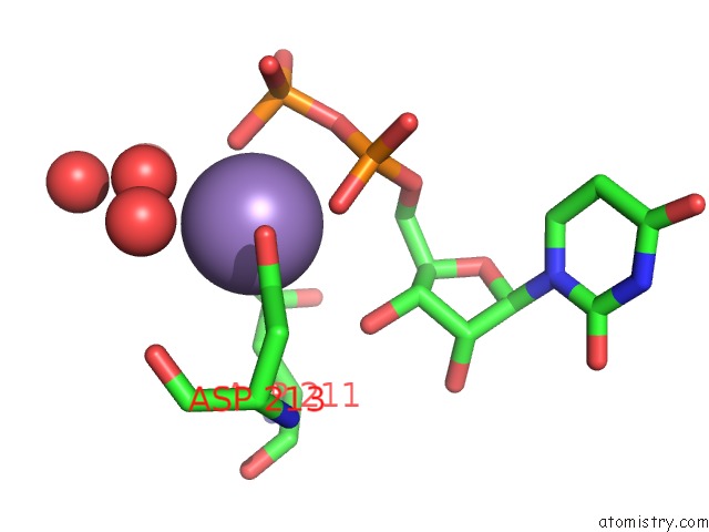
Mono view
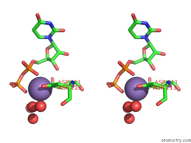
Stereo pair view
Mono view
Stereo pair view
A full contact list of Manganese with other atoms in the Mn binding
site number 1 of Glycosyltransferase A + Udp + H Antigen Acceptor within 5.0Å range:
|
Reference:
S.I.Patenaude,
N.O.Seto,
S.N.Borisova,
A.Szpacenko,
S.L.Marcus,
M.M.Palcic,
S.V.Evans.
The Structural Basis For Specificity in Human Abo(H) Blood Group Biosynthesis. Nat.Struct.Biol. V. 9 685 2002.
ISSN: ISSN 1072-8368
PubMed: 12198488
DOI: 10.1038/NSB832
Page generated: Sat Oct 5 11:37:08 2024
ISSN: ISSN 1072-8368
PubMed: 12198488
DOI: 10.1038/NSB832
Last articles
Ca in 5NRMCa in 5NUC
Ca in 5NRT
Ca in 5NSA
Ca in 5NPF
Ca in 5NQ2
Ca in 5N97
Ca in 5N8P
Ca in 5NMR
Ca in 5NN9