Manganese »
PDB 1khe-1lte »
1lt1 »
Manganese in PDB 1lt1: Sliding Helix Induced Change of Coordination Geometry in A Model Di-Mn(II) Protein
Protein crystallography data
The structure of Sliding Helix Induced Change of Coordination Geometry in A Model Di-Mn(II) Protein, PDB code: 1lt1
was solved by
L.Di Costanzo,
S.Geremia,
with X-Ray Crystallography technique. A brief refinement statistics is given in the table below:
| Resolution Low / High (Å) | 20.00 / 1.91 |
| Space group | P 21 21 21 |
| Cell size a, b, c (Å), α, β, γ (°) | 38.225, 89.270, 146.288, 90.00, 90.00, 90.00 |
| R / Rfree (%) | 20.1 / 24.5 |
Manganese Binding Sites:
Pages:
>>> Page 1 <<< Page 2, Binding sites: 11 - 12;Binding sites:
The binding sites of Manganese atom in the Sliding Helix Induced Change of Coordination Geometry in A Model Di-Mn(II) Protein (pdb code 1lt1). This binding sites where shown within 5.0 Angstroms radius around Manganese atom.In total 12 binding sites of Manganese where determined in the Sliding Helix Induced Change of Coordination Geometry in A Model Di-Mn(II) Protein, PDB code: 1lt1:
Jump to Manganese binding site number: 1; 2; 3; 4; 5; 6; 7; 8; 9; 10;
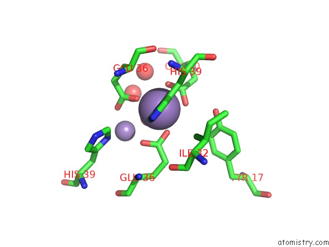
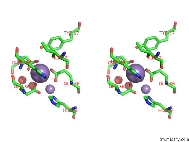
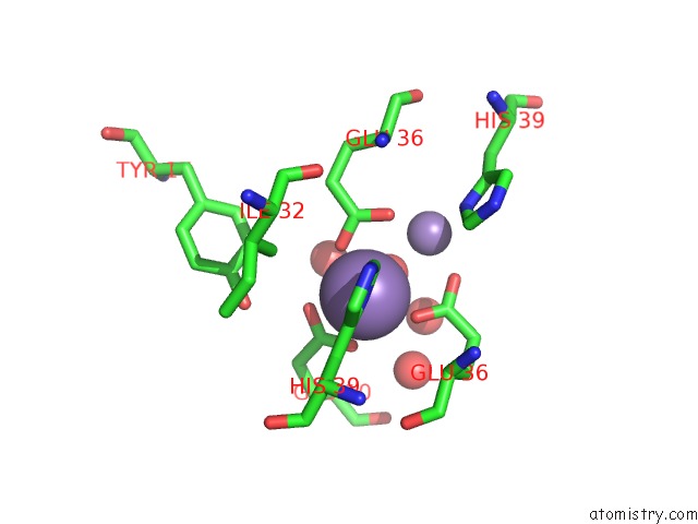
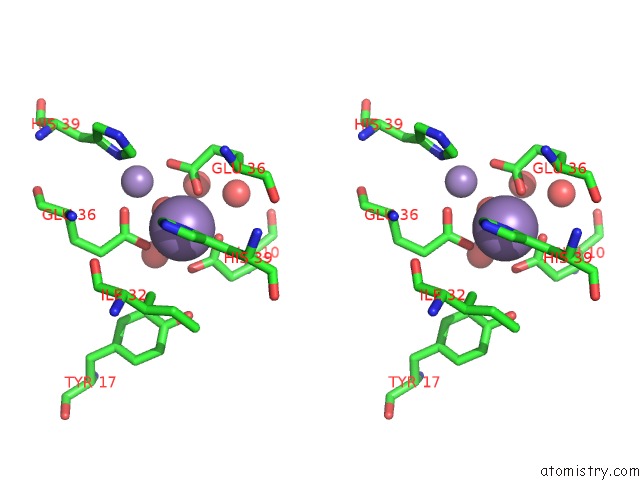
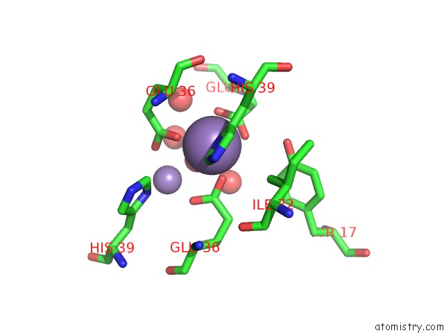
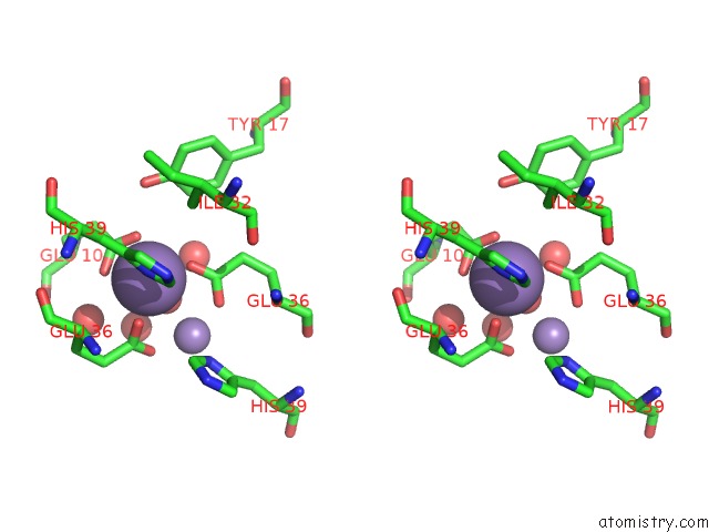
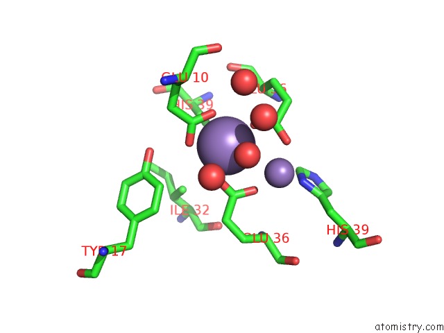
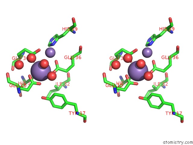
Manganese binding site 1 out of 12 in 1lt1
Go back to
Manganese binding site 1 out
of 12 in the Sliding Helix Induced Change of Coordination Geometry in A Model Di-Mn(II) Protein
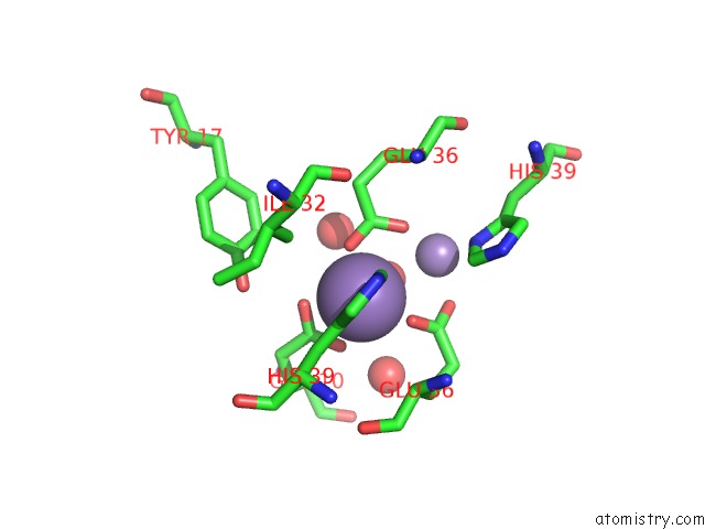
Mono view
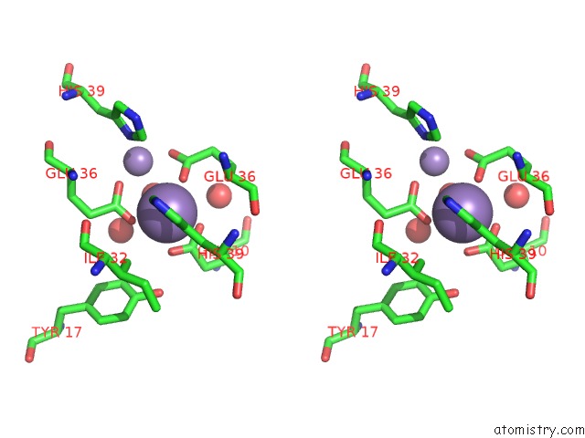
Stereo pair view
Mono view
Stereo pair view
A full contact list of Manganese with other atoms in the Mn binding
site number 1 of Sliding Helix Induced Change of Coordination Geometry in A Model Di-Mn(II) Protein within 5.0Å range:
|
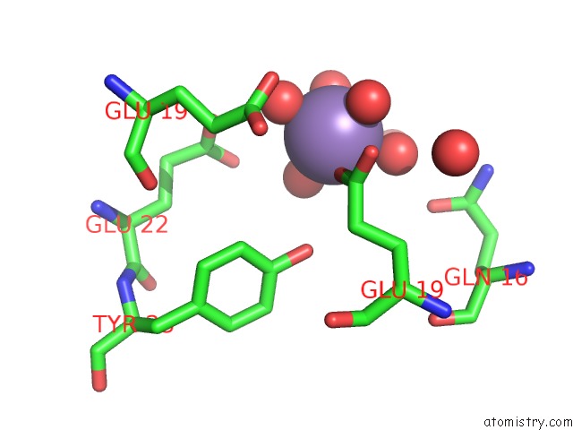
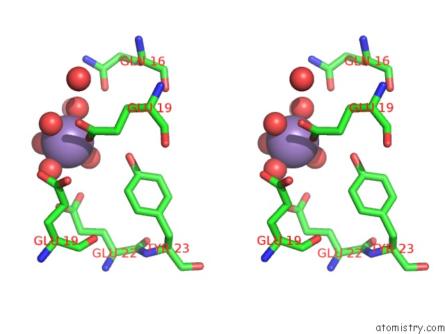
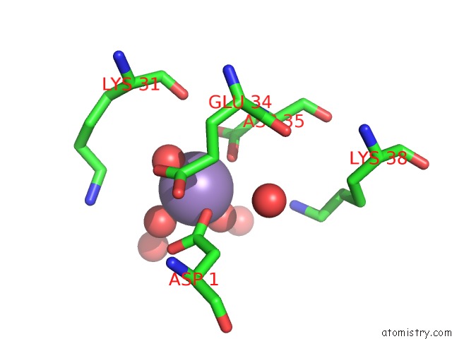
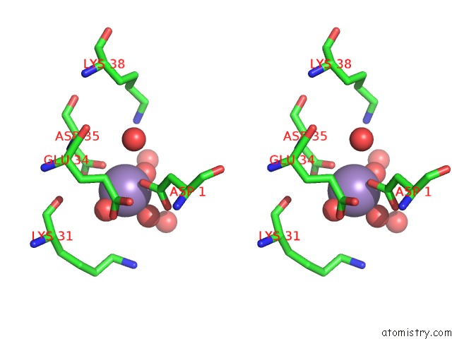
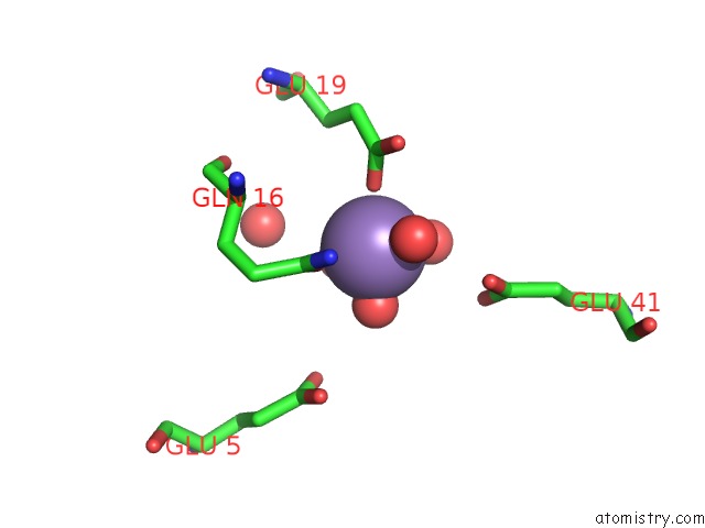
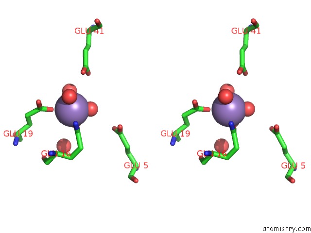
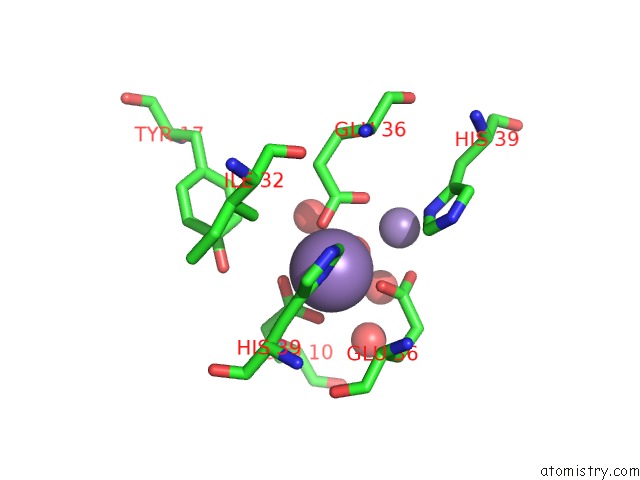
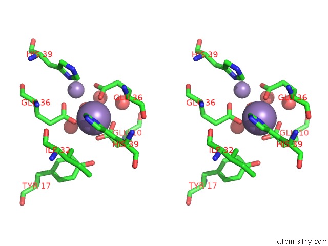
Manganese binding site 2 out of 12 in 1lt1
Go back to
Manganese binding site 2 out
of 12 in the Sliding Helix Induced Change of Coordination Geometry in A Model Di-Mn(II) Protein
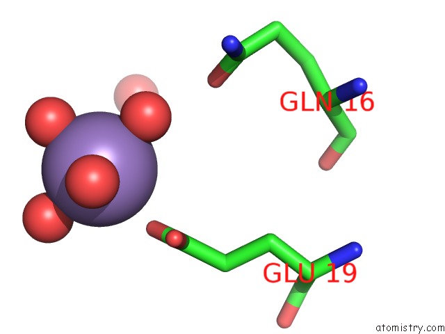
Mono view
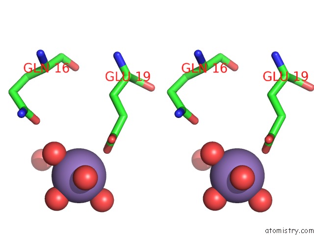
Stereo pair view
Mono view
Stereo pair view
A full contact list of Manganese with other atoms in the Mn binding
site number 2 of Sliding Helix Induced Change of Coordination Geometry in A Model Di-Mn(II) Protein within 5.0Å range:
|
Manganese binding site 3 out of 12 in 1lt1
Go back to
Manganese binding site 3 out
of 12 in the Sliding Helix Induced Change of Coordination Geometry in A Model Di-Mn(II) Protein
Mono view
Stereo pair view
Mono view
Stereo pair view
A full contact list of Manganese with other atoms in the Mn binding
site number 3 of Sliding Helix Induced Change of Coordination Geometry in A Model Di-Mn(II) Protein within 5.0Å range:
|
Manganese binding site 4 out of 12 in 1lt1
Go back to
Manganese binding site 4 out
of 12 in the Sliding Helix Induced Change of Coordination Geometry in A Model Di-Mn(II) Protein
Mono view
Stereo pair view
Mono view
Stereo pair view
A full contact list of Manganese with other atoms in the Mn binding
site number 4 of Sliding Helix Induced Change of Coordination Geometry in A Model Di-Mn(II) Protein within 5.0Å range:
|
Manganese binding site 5 out of 12 in 1lt1
Go back to
Manganese binding site 5 out
of 12 in the Sliding Helix Induced Change of Coordination Geometry in A Model Di-Mn(II) Protein
Mono view
Stereo pair view
Mono view
Stereo pair view
A full contact list of Manganese with other atoms in the Mn binding
site number 5 of Sliding Helix Induced Change of Coordination Geometry in A Model Di-Mn(II) Protein within 5.0Å range:
|
Manganese binding site 6 out of 12 in 1lt1
Go back to
Manganese binding site 6 out
of 12 in the Sliding Helix Induced Change of Coordination Geometry in A Model Di-Mn(II) Protein
Mono view
Stereo pair view
Mono view
Stereo pair view
A full contact list of Manganese with other atoms in the Mn binding
site number 6 of Sliding Helix Induced Change of Coordination Geometry in A Model Di-Mn(II) Protein within 5.0Å range:
|
Manganese binding site 7 out of 12 in 1lt1
Go back to
Manganese binding site 7 out
of 12 in the Sliding Helix Induced Change of Coordination Geometry in A Model Di-Mn(II) Protein
Mono view
Stereo pair view
Mono view
Stereo pair view
A full contact list of Manganese with other atoms in the Mn binding
site number 7 of Sliding Helix Induced Change of Coordination Geometry in A Model Di-Mn(II) Protein within 5.0Å range:
|
Manganese binding site 8 out of 12 in 1lt1
Go back to
Manganese binding site 8 out
of 12 in the Sliding Helix Induced Change of Coordination Geometry in A Model Di-Mn(II) Protein
Mono view
Stereo pair view
Mono view
Stereo pair view
A full contact list of Manganese with other atoms in the Mn binding
site number 8 of Sliding Helix Induced Change of Coordination Geometry in A Model Di-Mn(II) Protein within 5.0Å range:
|
Manganese binding site 9 out of 12 in 1lt1
Go back to
Manganese binding site 9 out
of 12 in the Sliding Helix Induced Change of Coordination Geometry in A Model Di-Mn(II) Protein
Mono view
Stereo pair view
Mono view
Stereo pair view
A full contact list of Manganese with other atoms in the Mn binding
site number 9 of Sliding Helix Induced Change of Coordination Geometry in A Model Di-Mn(II) Protein within 5.0Å range:
|
Manganese binding site 10 out of 12 in 1lt1
Go back to
Manganese binding site 10 out
of 12 in the Sliding Helix Induced Change of Coordination Geometry in A Model Di-Mn(II) Protein
Mono view
Stereo pair view
Mono view
Stereo pair view
A full contact list of Manganese with other atoms in the Mn binding
site number 10 of Sliding Helix Induced Change of Coordination Geometry in A Model Di-Mn(II) Protein within 5.0Å range:
|
Reference:
W.F.Degrado,
L.Di Costanzo,
S.Geremia,
A.Lombardi,
V.Pavone,
L.Randaccio.
Sliding Helix and Change of Coordination Geometry in A Model Di-Mnii Protein Angew.Chem.Int.Ed.Engl. V. 42 417 2003.
ISSN: ISSN 1433-7851
PubMed: 12569505
DOI: 10.1002/ANIE.200390127
Page generated: Sat Oct 5 11:34:23 2024
ISSN: ISSN 1433-7851
PubMed: 12569505
DOI: 10.1002/ANIE.200390127
Last articles
Zn in 9J0NZn in 9J0O
Zn in 9J0P
Zn in 9FJX
Zn in 9EKB
Zn in 9C0F
Zn in 9CAH
Zn in 9CH0
Zn in 9CH3
Zn in 9CH1