Manganese »
PDB 1j54-1khb »
1kgz »
Manganese in PDB 1kgz: Crystal Structure Analysis of the Anthranilate Phosphoribosyltransferase From Erwinia Carotovora (Current Name, Pectobacterium Carotovorum)
Enzymatic activity of Crystal Structure Analysis of the Anthranilate Phosphoribosyltransferase From Erwinia Carotovora (Current Name, Pectobacterium Carotovorum)
All present enzymatic activity of Crystal Structure Analysis of the Anthranilate Phosphoribosyltransferase From Erwinia Carotovora (Current Name, Pectobacterium Carotovorum):
2.4.2.18;
2.4.2.18;
Protein crystallography data
The structure of Crystal Structure Analysis of the Anthranilate Phosphoribosyltransferase From Erwinia Carotovora (Current Name, Pectobacterium Carotovorum), PDB code: 1kgz
was solved by
C.Kim,
N.-H.Xuong,
S.Edwards,
Madhusudan,
M.-C.Yee,
G.Spraggon,
S.E.Mills,
with X-Ray Crystallography technique. A brief refinement statistics is given in the table below:
| Resolution Low / High (Å) | 25.00 / 2.40 |
| Space group | P 21 21 2 |
| Cell size a, b, c (Å), α, β, γ (°) | 77.378, 81.686, 101.680, 90.00, 90.00, 90.00 |
| R / Rfree (%) | 21.4 / 26.5 |
Other elements in 1kgz:
The structure of Crystal Structure Analysis of the Anthranilate Phosphoribosyltransferase From Erwinia Carotovora (Current Name, Pectobacterium Carotovorum) also contains other interesting chemical elements:
| Mercury | (Hg) | 2 atoms |
Manganese Binding Sites:
The binding sites of Manganese atom in the Crystal Structure Analysis of the Anthranilate Phosphoribosyltransferase From Erwinia Carotovora (Current Name, Pectobacterium Carotovorum)
(pdb code 1kgz). This binding sites where shown within
5.0 Angstroms radius around Manganese atom.
In total 4 binding sites of Manganese where determined in the Crystal Structure Analysis of the Anthranilate Phosphoribosyltransferase From Erwinia Carotovora (Current Name, Pectobacterium Carotovorum), PDB code: 1kgz:
Jump to Manganese binding site number: 1; 2; 3; 4;
In total 4 binding sites of Manganese where determined in the Crystal Structure Analysis of the Anthranilate Phosphoribosyltransferase From Erwinia Carotovora (Current Name, Pectobacterium Carotovorum), PDB code: 1kgz:
Jump to Manganese binding site number: 1; 2; 3; 4;
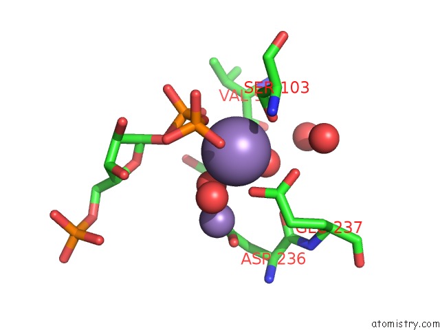
Manganese binding site 1 out of 4 in 1kgz
Go back to
Manganese binding site 1 out
of 4 in the Crystal Structure Analysis of the Anthranilate Phosphoribosyltransferase From Erwinia Carotovora (Current Name, Pectobacterium Carotovorum)
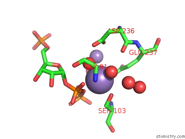
Mono view
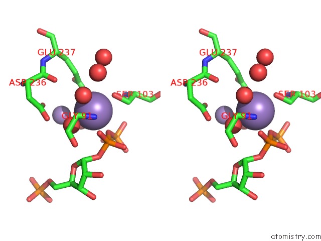
Stereo pair view
Mono view
Stereo pair view
A full contact list of Manganese with other atoms in the Mn binding
site number 1 of Crystal Structure Analysis of the Anthranilate Phosphoribosyltransferase From Erwinia Carotovora (Current Name, Pectobacterium Carotovorum) within 5.0Å range:
|
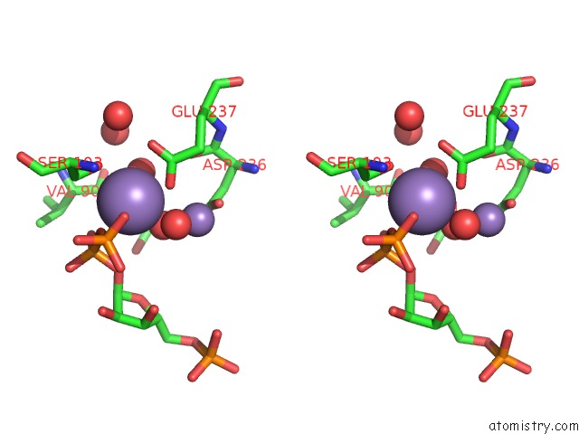
Manganese binding site 2 out of 4 in 1kgz
Go back to
Manganese binding site 2 out
of 4 in the Crystal Structure Analysis of the Anthranilate Phosphoribosyltransferase From Erwinia Carotovora (Current Name, Pectobacterium Carotovorum)
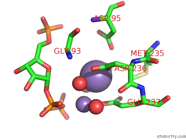
Mono view
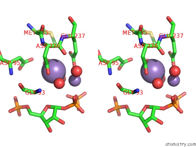
Stereo pair view
Mono view
Stereo pair view
A full contact list of Manganese with other atoms in the Mn binding
site number 2 of Crystal Structure Analysis of the Anthranilate Phosphoribosyltransferase From Erwinia Carotovora (Current Name, Pectobacterium Carotovorum) within 5.0Å range:
|
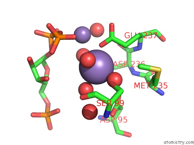
Manganese binding site 3 out of 4 in 1kgz
Go back to
Manganese binding site 3 out
of 4 in the Crystal Structure Analysis of the Anthranilate Phosphoribosyltransferase From Erwinia Carotovora (Current Name, Pectobacterium Carotovorum)
Mono view
Stereo pair view
Mono view
Stereo pair view
A full contact list of Manganese with other atoms in the Mn binding
site number 3 of Crystal Structure Analysis of the Anthranilate Phosphoribosyltransferase From Erwinia Carotovora (Current Name, Pectobacterium Carotovorum) within 5.0Å range:
|
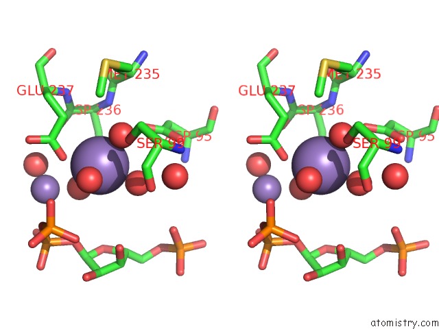
Manganese binding site 4 out of 4 in 1kgz
Go back to
Manganese binding site 4 out
of 4 in the Crystal Structure Analysis of the Anthranilate Phosphoribosyltransferase From Erwinia Carotovora (Current Name, Pectobacterium Carotovorum)
Mono view
Stereo pair view
Mono view
Stereo pair view
A full contact list of Manganese with other atoms in the Mn binding
site number 4 of Crystal Structure Analysis of the Anthranilate Phosphoribosyltransferase From Erwinia Carotovora (Current Name, Pectobacterium Carotovorum) within 5.0Å range:
|
Reference:
C.Kim,
N.-H.Xuong,
S.Edwards,
Madhusudan,
M.-C.Yee,
G.Spraggon,
S.E.Mills.
The Crystal Structure of Anthranilate Phosphoribosyltransferase From the Enterobacterium Pectobacterium Carotovorum Febs Lett. V. 523 239 2002.
ISSN: ISSN 0014-5793
PubMed: 12123839
DOI: 10.1016/S0014-5793(02)02905-8
Page generated: Sat Oct 5 11:20:23 2024
ISSN: ISSN 0014-5793
PubMed: 12123839
DOI: 10.1016/S0014-5793(02)02905-8
Last articles
Zn in 9J0NZn in 9J0O
Zn in 9J0P
Zn in 9FJX
Zn in 9EKB
Zn in 9C0F
Zn in 9CAH
Zn in 9CH0
Zn in 9CH3
Zn in 9CH1