Manganese »
PDB 1j54-1khb »
1kfl »
Manganese in PDB 1kfl: Crystal Structure of Phenylalanine-Regulated 3-Deoxy-D- Arabino-Heptulosonate-7-Phosphate Synthase (Dahp Synthase) From E.Coli Complexed with MN2+, Pep, and Phe
Enzymatic activity of Crystal Structure of Phenylalanine-Regulated 3-Deoxy-D- Arabino-Heptulosonate-7-Phosphate Synthase (Dahp Synthase) From E.Coli Complexed with MN2+, Pep, and Phe
All present enzymatic activity of Crystal Structure of Phenylalanine-Regulated 3-Deoxy-D- Arabino-Heptulosonate-7-Phosphate Synthase (Dahp Synthase) From E.Coli Complexed with MN2+, Pep, and Phe:
4.1.2.15;
4.1.2.15;
Protein crystallography data
The structure of Crystal Structure of Phenylalanine-Regulated 3-Deoxy-D- Arabino-Heptulosonate-7-Phosphate Synthase (Dahp Synthase) From E.Coli Complexed with MN2+, Pep, and Phe, PDB code: 1kfl
was solved by
I.A.Shumilin,
C.Zhao,
R.Bauerle,
R.H.Kretsinger,
with X-Ray Crystallography technique. A brief refinement statistics is given in the table below:
| Resolution Low / High (Å) | 20.00 / 2.80 |
| Space group | C 1 2 1 |
| Cell size a, b, c (Å), α, β, γ (°) | 290.097, 90.097, 155.798, 90.00, 120.77, 90.00 |
| R / Rfree (%) | 21.8 / 24.6 |
Manganese Binding Sites:
The binding sites of Manganese atom in the Crystal Structure of Phenylalanine-Regulated 3-Deoxy-D- Arabino-Heptulosonate-7-Phosphate Synthase (Dahp Synthase) From E.Coli Complexed with MN2+, Pep, and Phe
(pdb code 1kfl). This binding sites where shown within
5.0 Angstroms radius around Manganese atom.
In total 8 binding sites of Manganese where determined in the Crystal Structure of Phenylalanine-Regulated 3-Deoxy-D- Arabino-Heptulosonate-7-Phosphate Synthase (Dahp Synthase) From E.Coli Complexed with MN2+, Pep, and Phe, PDB code: 1kfl:
Jump to Manganese binding site number: 1; 2; 3; 4; 5; 6; 7; 8;
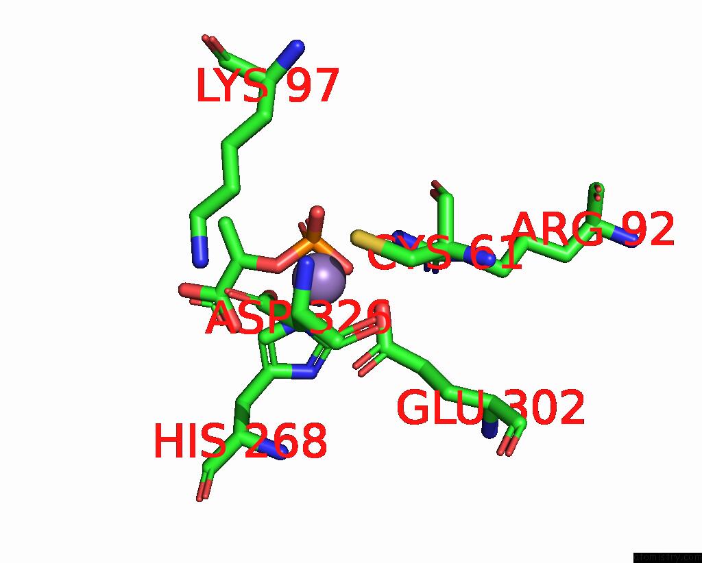
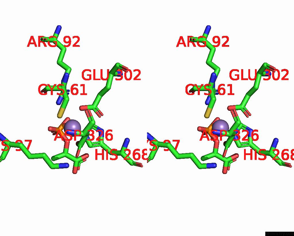
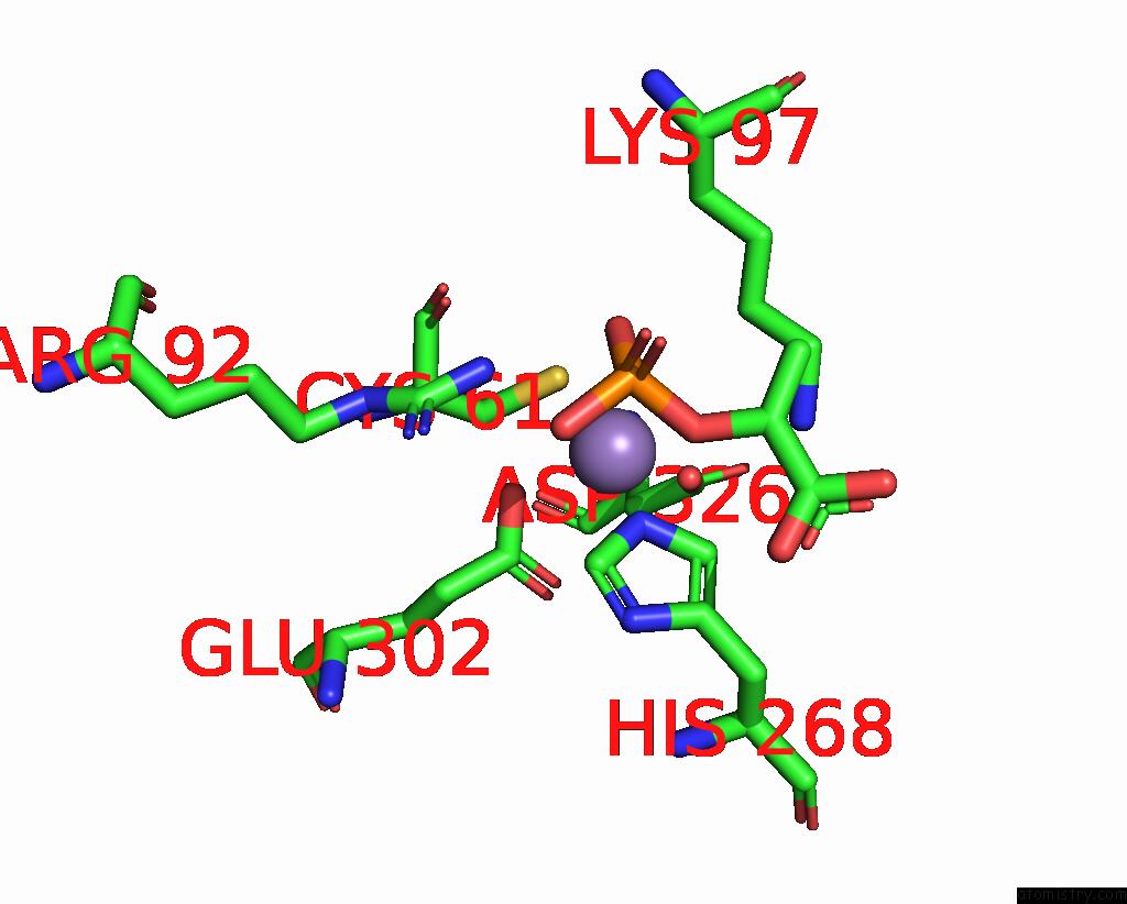
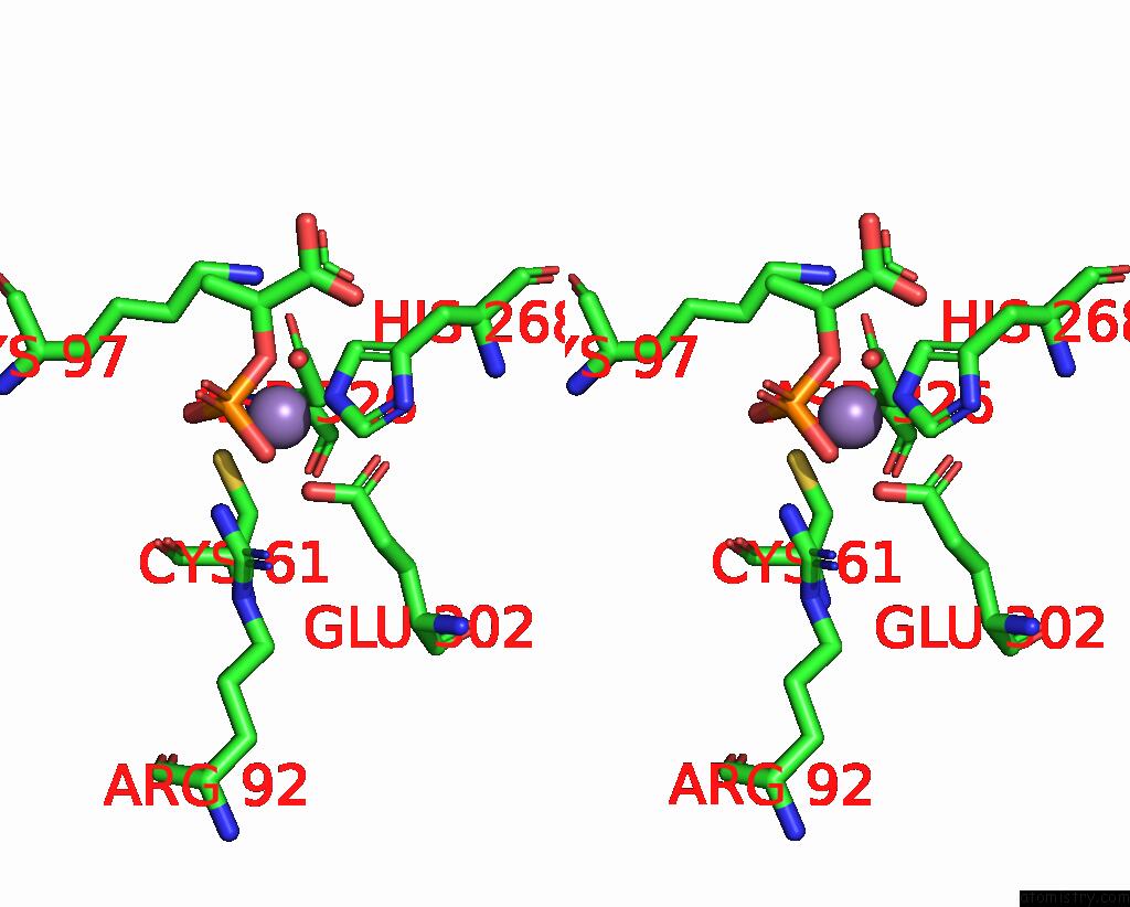
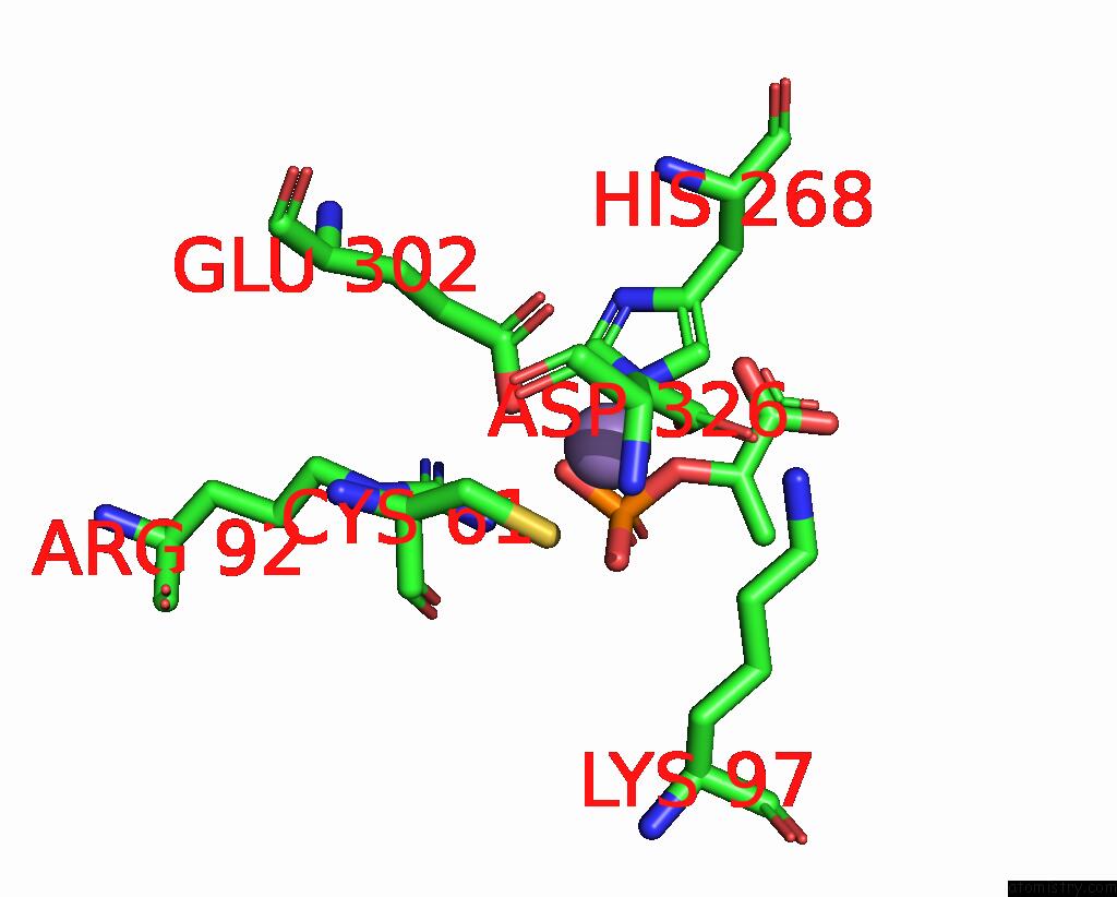
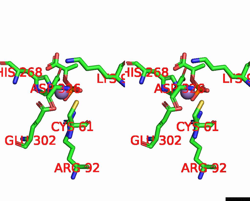
Manganese binding site 1 out of 8 in 1kfl
Go back to
Manganese binding site 1 out
of 8 in the Crystal Structure of Phenylalanine-Regulated 3-Deoxy-D- Arabino-Heptulosonate-7-Phosphate Synthase (Dahp Synthase) From E.Coli Complexed with MN2+, Pep, and Phe
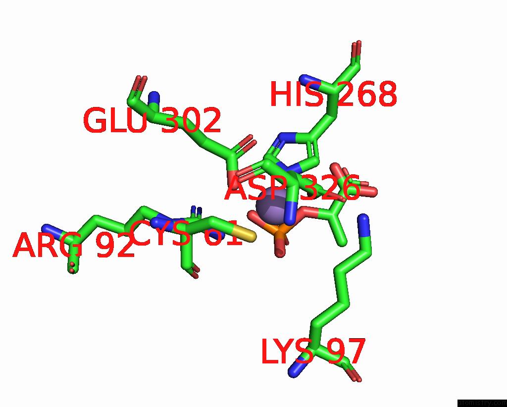
Mono view
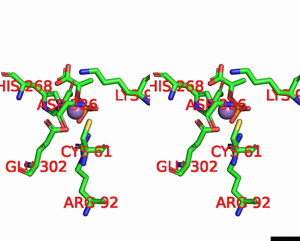
Stereo pair view
Mono view
Stereo pair view
A full contact list of Manganese with other atoms in the Mn binding
site number 1 of Crystal Structure of Phenylalanine-Regulated 3-Deoxy-D- Arabino-Heptulosonate-7-Phosphate Synthase (Dahp Synthase) From E.Coli Complexed with MN2+, Pep, and Phe within 5.0Å range:
|
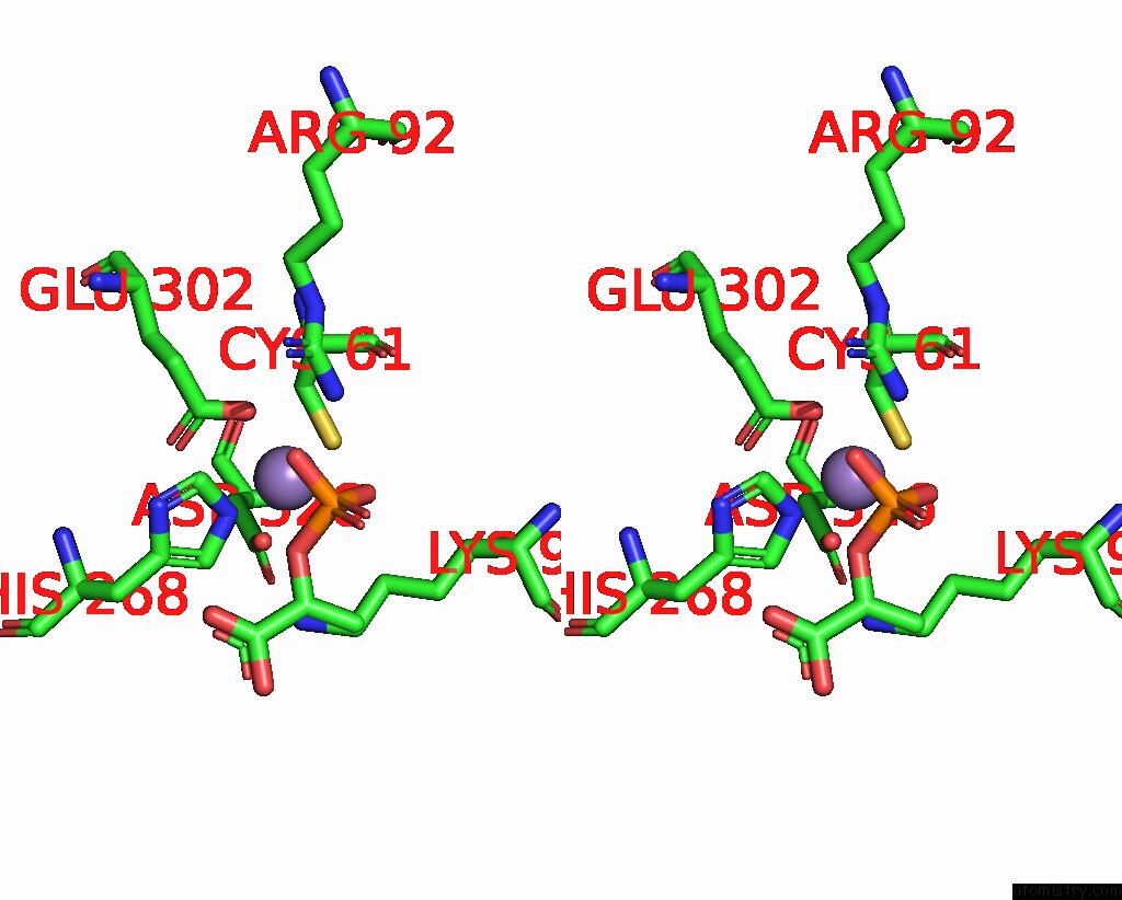
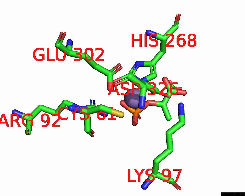
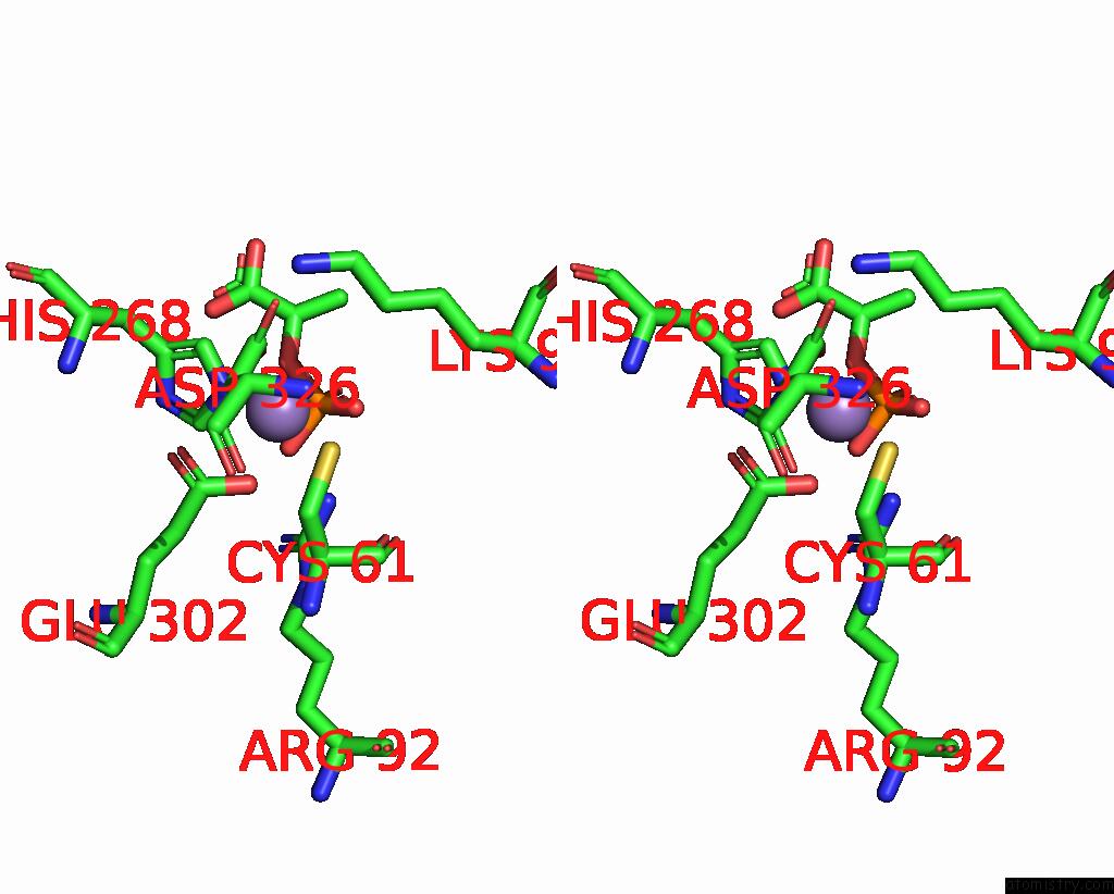
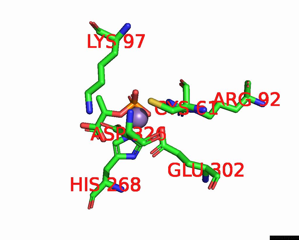
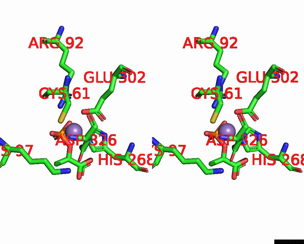
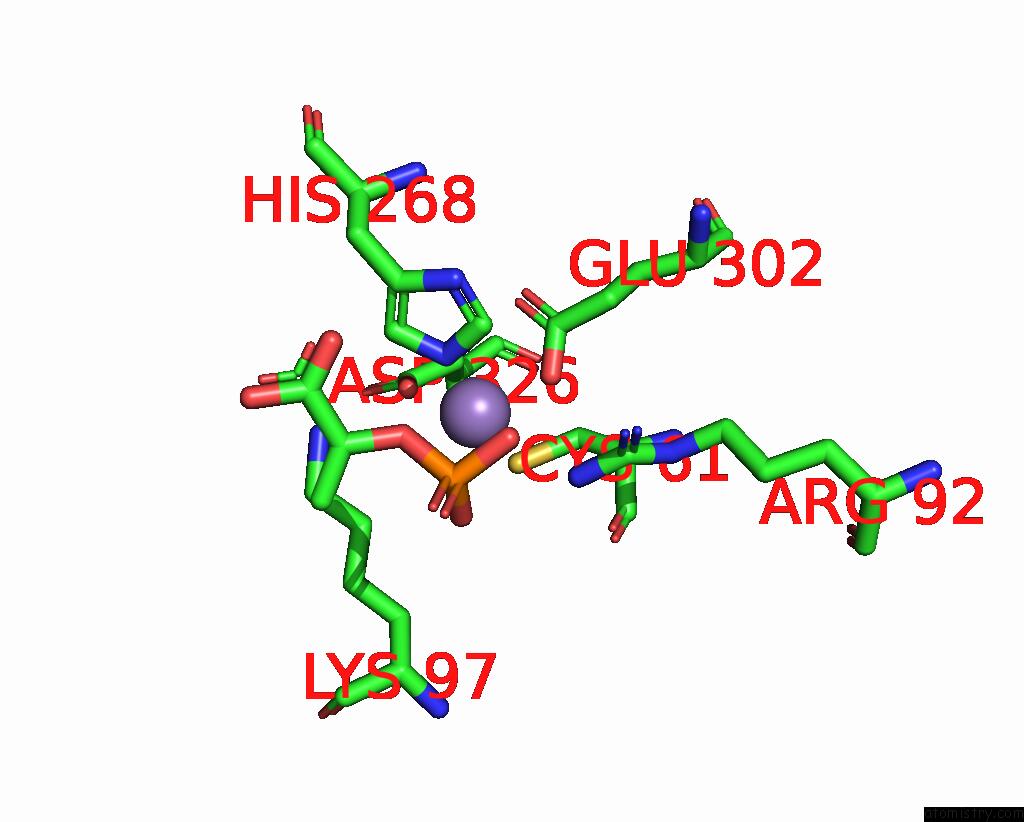
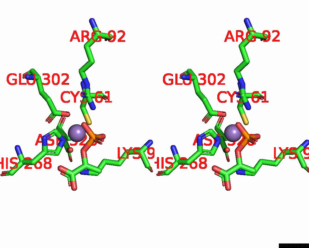
Manganese binding site 2 out of 8 in 1kfl
Go back to
Manganese binding site 2 out
of 8 in the Crystal Structure of Phenylalanine-Regulated 3-Deoxy-D- Arabino-Heptulosonate-7-Phosphate Synthase (Dahp Synthase) From E.Coli Complexed with MN2+, Pep, and Phe
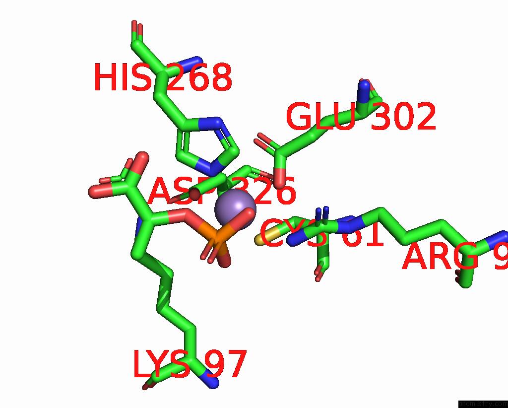
Mono view
Stereo pair view
Mono view
Stereo pair view
A full contact list of Manganese with other atoms in the Mn binding
site number 2 of Crystal Structure of Phenylalanine-Regulated 3-Deoxy-D- Arabino-Heptulosonate-7-Phosphate Synthase (Dahp Synthase) From E.Coli Complexed with MN2+, Pep, and Phe within 5.0Å range:
|
Manganese binding site 3 out of 8 in 1kfl
Go back to
Manganese binding site 3 out
of 8 in the Crystal Structure of Phenylalanine-Regulated 3-Deoxy-D- Arabino-Heptulosonate-7-Phosphate Synthase (Dahp Synthase) From E.Coli Complexed with MN2+, Pep, and Phe
Mono view
Stereo pair view
Mono view
Stereo pair view
A full contact list of Manganese with other atoms in the Mn binding
site number 3 of Crystal Structure of Phenylalanine-Regulated 3-Deoxy-D- Arabino-Heptulosonate-7-Phosphate Synthase (Dahp Synthase) From E.Coli Complexed with MN2+, Pep, and Phe within 5.0Å range:
|
Manganese binding site 4 out of 8 in 1kfl
Go back to
Manganese binding site 4 out
of 8 in the Crystal Structure of Phenylalanine-Regulated 3-Deoxy-D- Arabino-Heptulosonate-7-Phosphate Synthase (Dahp Synthase) From E.Coli Complexed with MN2+, Pep, and Phe
Mono view
Stereo pair view
Mono view
Stereo pair view
A full contact list of Manganese with other atoms in the Mn binding
site number 4 of Crystal Structure of Phenylalanine-Regulated 3-Deoxy-D- Arabino-Heptulosonate-7-Phosphate Synthase (Dahp Synthase) From E.Coli Complexed with MN2+, Pep, and Phe within 5.0Å range:
|
Manganese binding site 5 out of 8 in 1kfl
Go back to
Manganese binding site 5 out
of 8 in the Crystal Structure of Phenylalanine-Regulated 3-Deoxy-D- Arabino-Heptulosonate-7-Phosphate Synthase (Dahp Synthase) From E.Coli Complexed with MN2+, Pep, and Phe
Mono view
Stereo pair view
Mono view
Stereo pair view
A full contact list of Manganese with other atoms in the Mn binding
site number 5 of Crystal Structure of Phenylalanine-Regulated 3-Deoxy-D- Arabino-Heptulosonate-7-Phosphate Synthase (Dahp Synthase) From E.Coli Complexed with MN2+, Pep, and Phe within 5.0Å range:
|
Manganese binding site 6 out of 8 in 1kfl
Go back to
Manganese binding site 6 out
of 8 in the Crystal Structure of Phenylalanine-Regulated 3-Deoxy-D- Arabino-Heptulosonate-7-Phosphate Synthase (Dahp Synthase) From E.Coli Complexed with MN2+, Pep, and Phe
Mono view
Stereo pair view
Mono view
Stereo pair view
A full contact list of Manganese with other atoms in the Mn binding
site number 6 of Crystal Structure of Phenylalanine-Regulated 3-Deoxy-D- Arabino-Heptulosonate-7-Phosphate Synthase (Dahp Synthase) From E.Coli Complexed with MN2+, Pep, and Phe within 5.0Å range:
|
Manganese binding site 7 out of 8 in 1kfl
Go back to
Manganese binding site 7 out
of 8 in the Crystal Structure of Phenylalanine-Regulated 3-Deoxy-D- Arabino-Heptulosonate-7-Phosphate Synthase (Dahp Synthase) From E.Coli Complexed with MN2+, Pep, and Phe
Mono view
Stereo pair view
Mono view
Stereo pair view
A full contact list of Manganese with other atoms in the Mn binding
site number 7 of Crystal Structure of Phenylalanine-Regulated 3-Deoxy-D- Arabino-Heptulosonate-7-Phosphate Synthase (Dahp Synthase) From E.Coli Complexed with MN2+, Pep, and Phe within 5.0Å range:
|
Manganese binding site 8 out of 8 in 1kfl
Go back to
Manganese binding site 8 out
of 8 in the Crystal Structure of Phenylalanine-Regulated 3-Deoxy-D- Arabino-Heptulosonate-7-Phosphate Synthase (Dahp Synthase) From E.Coli Complexed with MN2+, Pep, and Phe
Mono view
Stereo pair view
Mono view
Stereo pair view
A full contact list of Manganese with other atoms in the Mn binding
site number 8 of Crystal Structure of Phenylalanine-Regulated 3-Deoxy-D- Arabino-Heptulosonate-7-Phosphate Synthase (Dahp Synthase) From E.Coli Complexed with MN2+, Pep, and Phe within 5.0Å range:
|
Reference:
I.A.Shumilin,
C.Zhao,
R.Bauerle,
R.H.Kretsinger.
Allosteric Inhibition of 3-Deoxy-D-Arabino-Heptulosonate-7-Phosphate Synthase Alters the Coordination of Both Substrates. J.Mol.Biol. V. 320 1147 2002.
ISSN: ISSN 0022-2836
PubMed: 12126632
DOI: 10.1016/S0022-2836(02)00545-4
Page generated: Sat Oct 5 11:20:16 2024
ISSN: ISSN 0022-2836
PubMed: 12126632
DOI: 10.1016/S0022-2836(02)00545-4
Last articles
Ca in 5NERCa in 5NEM
Ca in 5NE5
Ca in 5NBP
Ca in 5NBN
Ca in 5NBM
Ca in 5NBL
Ca in 5N7G
Ca in 5N7F
Ca in 5N7D