Manganese »
PDB 1ho5-1j53 »
1it6 »
Manganese in PDB 1it6: Crystal Structure of the Complex Between Calyculin A and the Catalytic Subunit of Protein Phosphatase 1
Enzymatic activity of Crystal Structure of the Complex Between Calyculin A and the Catalytic Subunit of Protein Phosphatase 1
All present enzymatic activity of Crystal Structure of the Complex Between Calyculin A and the Catalytic Subunit of Protein Phosphatase 1:
3.1.3.16;
3.1.3.16;
Protein crystallography data
The structure of Crystal Structure of the Complex Between Calyculin A and the Catalytic Subunit of Protein Phosphatase 1, PDB code: 1it6
was solved by
A.Kita,
S.Matsunaga,
A.Takai,
H.Kataiwa,
T.Wakimoto,
N.Fusetani,
M.Isobe,
K.Miki,
with X-Ray Crystallography technique. A brief refinement statistics is given in the table below:
| Resolution Low / High (Å) | 20.00 / 2.00 |
| Space group | P 21 21 2 |
| Cell size a, b, c (Å), α, β, γ (°) | 98.090, 136.360, 61.580, 90.00, 90.00, 90.00 |
| R / Rfree (%) | 18.2 / 21.8 |
Manganese Binding Sites:
The binding sites of Manganese atom in the Crystal Structure of the Complex Between Calyculin A and the Catalytic Subunit of Protein Phosphatase 1
(pdb code 1it6). This binding sites where shown within
5.0 Angstroms radius around Manganese atom.
In total 4 binding sites of Manganese where determined in the Crystal Structure of the Complex Between Calyculin A and the Catalytic Subunit of Protein Phosphatase 1, PDB code: 1it6:
Jump to Manganese binding site number: 1; 2; 3; 4;
In total 4 binding sites of Manganese where determined in the Crystal Structure of the Complex Between Calyculin A and the Catalytic Subunit of Protein Phosphatase 1, PDB code: 1it6:
Jump to Manganese binding site number: 1; 2; 3; 4;
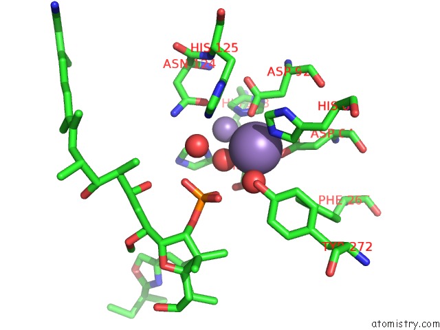
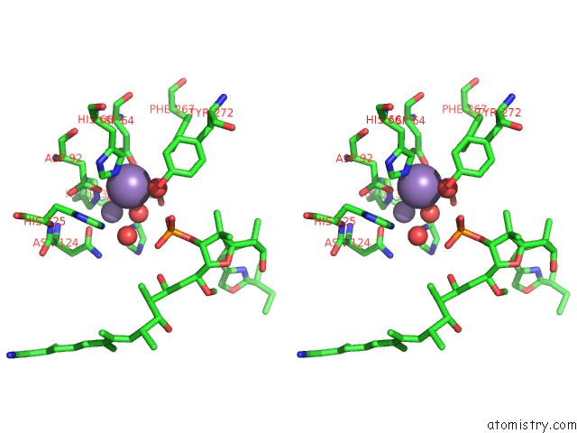
Manganese binding site 1 out of 4 in 1it6
Go back to
Manganese binding site 1 out
of 4 in the Crystal Structure of the Complex Between Calyculin A and the Catalytic Subunit of Protein Phosphatase 1
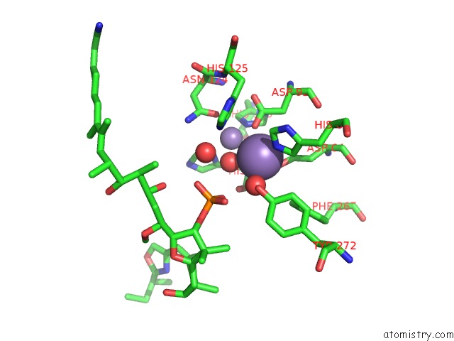
Mono view
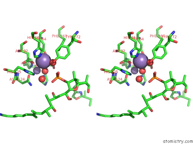
Stereo pair view
Mono view
Stereo pair view
A full contact list of Manganese with other atoms in the Mn binding
site number 1 of Crystal Structure of the Complex Between Calyculin A and the Catalytic Subunit of Protein Phosphatase 1 within 5.0Å range:
|
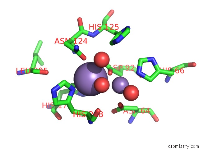
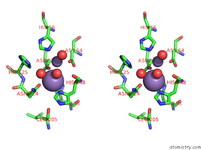
Manganese binding site 2 out of 4 in 1it6
Go back to
Manganese binding site 2 out
of 4 in the Crystal Structure of the Complex Between Calyculin A and the Catalytic Subunit of Protein Phosphatase 1
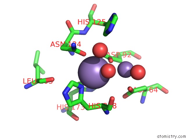
Mono view
Stereo pair view
Mono view
Stereo pair view
A full contact list of Manganese with other atoms in the Mn binding
site number 2 of Crystal Structure of the Complex Between Calyculin A and the Catalytic Subunit of Protein Phosphatase 1 within 5.0Å range:
|
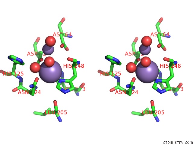
Manganese binding site 3 out of 4 in 1it6
Go back to
Manganese binding site 3 out
of 4 in the Crystal Structure of the Complex Between Calyculin A and the Catalytic Subunit of Protein Phosphatase 1
Mono view
Stereo pair view
Mono view
Stereo pair view
A full contact list of Manganese with other atoms in the Mn binding
site number 3 of Crystal Structure of the Complex Between Calyculin A and the Catalytic Subunit of Protein Phosphatase 1 within 5.0Å range:
|
Manganese binding site 4 out of 4 in 1it6
Go back to
Manganese binding site 4 out
of 4 in the Crystal Structure of the Complex Between Calyculin A and the Catalytic Subunit of Protein Phosphatase 1
Mono view
Stereo pair view
Mono view
Stereo pair view
A full contact list of Manganese with other atoms in the Mn binding
site number 4 of Crystal Structure of the Complex Between Calyculin A and the Catalytic Subunit of Protein Phosphatase 1 within 5.0Å range:
|
Reference:
A.Kita,
S.Matsunaga,
A.Takai,
H.Kataiwa,
T.Wakimoto,
N.Fusetani,
M.Isobe,
K.Miki.
Crystal Structure of the Complex Between Calyculin A and the Catalytic Subunit of Protein Phosphatase 1. Structure V. 10 715 2002.
ISSN: ISSN 0969-2126
PubMed: 12015153
DOI: 10.1016/S0969-2126(02)00764-5
Page generated: Sat Oct 5 11:03:46 2024
ISSN: ISSN 0969-2126
PubMed: 12015153
DOI: 10.1016/S0969-2126(02)00764-5
Last articles
Ca in 5N5PCa in 5N6N
Ca in 5N5K
Ca in 5N5W
Ca in 5N5J
Ca in 5N4L
Ca in 5N3Y
Ca in 5N31
Ca in 5N3V
Ca in 5N34