Manganese »
PDB 1ho5-1j53 »
1i08 »
Manganese in PDB 1i08: Crystal Structure Analysis of the H30A Mutant of Manganese Superoxide Dismutase From E. Coli
Enzymatic activity of Crystal Structure Analysis of the H30A Mutant of Manganese Superoxide Dismutase From E. Coli
All present enzymatic activity of Crystal Structure Analysis of the H30A Mutant of Manganese Superoxide Dismutase From E. Coli:
1.15.1.1;
1.15.1.1;
Protein crystallography data
The structure of Crystal Structure Analysis of the H30A Mutant of Manganese Superoxide Dismutase From E. Coli, PDB code: 1i08
was solved by
R.A.Edwards,
M.M.Whittaker,
J.W.Whittaker,
E.N.Baker,
G.B.Jameson,
with X-Ray Crystallography technique. A brief refinement statistics is given in the table below:
| Resolution Low / High (Å) | 40.00 / 2.20 |
| Space group | C 2 2 21 |
| Cell size a, b, c (Å), α, β, γ (°) | 100.681, 109.110, 181.072, 90.00, 90.00, 90.00 |
| R / Rfree (%) | 18.4 / 21.1 |
Manganese Binding Sites:
The binding sites of Manganese atom in the Crystal Structure Analysis of the H30A Mutant of Manganese Superoxide Dismutase From E. Coli
(pdb code 1i08). This binding sites where shown within
5.0 Angstroms radius around Manganese atom.
In total 4 binding sites of Manganese where determined in the Crystal Structure Analysis of the H30A Mutant of Manganese Superoxide Dismutase From E. Coli, PDB code: 1i08:
Jump to Manganese binding site number: 1; 2; 3; 4;
In total 4 binding sites of Manganese where determined in the Crystal Structure Analysis of the H30A Mutant of Manganese Superoxide Dismutase From E. Coli, PDB code: 1i08:
Jump to Manganese binding site number: 1; 2; 3; 4;
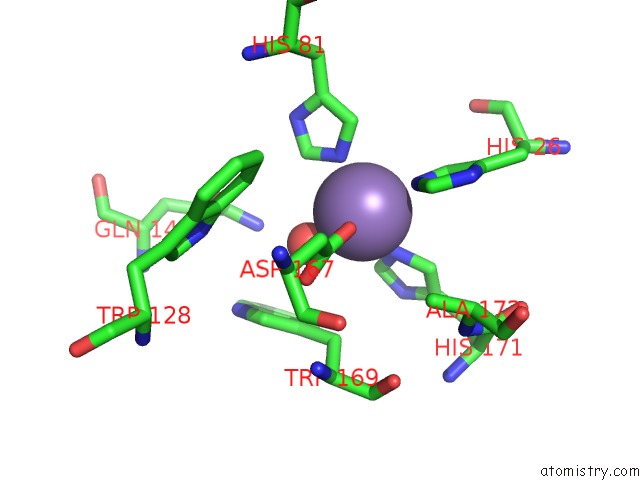
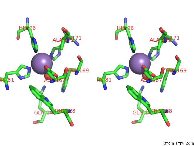
Manganese binding site 1 out of 4 in 1i08
Go back to
Manganese binding site 1 out
of 4 in the Crystal Structure Analysis of the H30A Mutant of Manganese Superoxide Dismutase From E. Coli
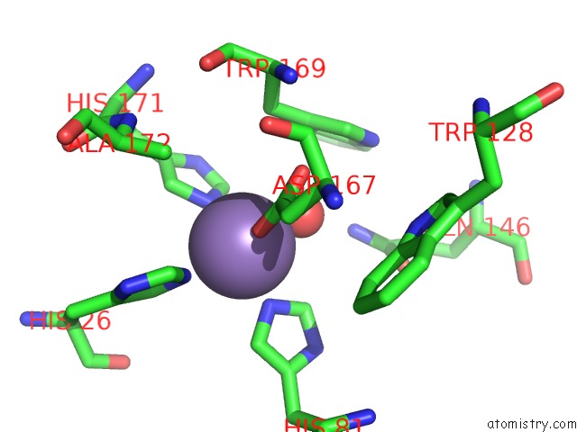
Mono view
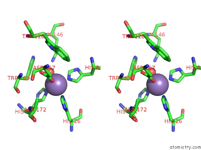
Stereo pair view
Mono view
Stereo pair view
A full contact list of Manganese with other atoms in the Mn binding
site number 1 of Crystal Structure Analysis of the H30A Mutant of Manganese Superoxide Dismutase From E. Coli within 5.0Å range:
|
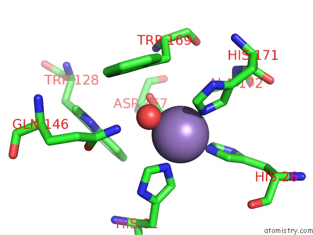
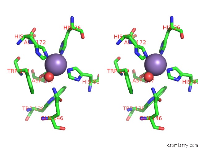
Manganese binding site 2 out of 4 in 1i08
Go back to
Manganese binding site 2 out
of 4 in the Crystal Structure Analysis of the H30A Mutant of Manganese Superoxide Dismutase From E. Coli
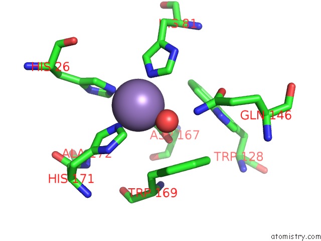
Mono view
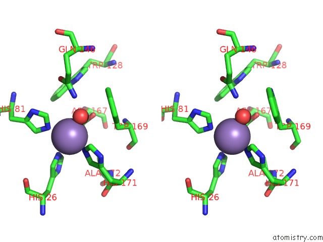
Stereo pair view
Mono view
Stereo pair view
A full contact list of Manganese with other atoms in the Mn binding
site number 2 of Crystal Structure Analysis of the H30A Mutant of Manganese Superoxide Dismutase From E. Coli within 5.0Å range:
|
Manganese binding site 3 out of 4 in 1i08
Go back to
Manganese binding site 3 out
of 4 in the Crystal Structure Analysis of the H30A Mutant of Manganese Superoxide Dismutase From E. Coli
Mono view
Stereo pair view
Mono view
Stereo pair view
A full contact list of Manganese with other atoms in the Mn binding
site number 3 of Crystal Structure Analysis of the H30A Mutant of Manganese Superoxide Dismutase From E. Coli within 5.0Å range:
|
Manganese binding site 4 out of 4 in 1i08
Go back to
Manganese binding site 4 out
of 4 in the Crystal Structure Analysis of the H30A Mutant of Manganese Superoxide Dismutase From E. Coli
Mono view
Stereo pair view
Mono view
Stereo pair view
A full contact list of Manganese with other atoms in the Mn binding
site number 4 of Crystal Structure Analysis of the H30A Mutant of Manganese Superoxide Dismutase From E. Coli within 5.0Å range:
|
Reference:
R.A.Edwards,
M.M.Whittaker,
J.W.Whittaker,
E.N.Baker,
G.B.Jameson.
Removing A Hydrogen Bond in the Dimer Interface of Escherichia Coli Manganese Superoxide Dismutase Alters Structure and Reactivity. Biochemistry V. 40 4622 2001.
ISSN: ISSN 0006-2960
PubMed: 11294629
DOI: 10.1021/BI002403H
Page generated: Sat Oct 5 10:58:19 2024
ISSN: ISSN 0006-2960
PubMed: 11294629
DOI: 10.1021/BI002403H
Last articles
Zn in 9J0NZn in 9J0O
Zn in 9J0P
Zn in 9FJX
Zn in 9EKB
Zn in 9C0F
Zn in 9CAH
Zn in 9CH0
Zn in 9CH3
Zn in 9CH1