Manganese »
PDB 1ho5-1j53 »
1hqw »
Manganese in PDB 1hqw: Crystal Structure of the Complex of Concanavalin A with A Tripeptide Ypy
Protein crystallography data
The structure of Crystal Structure of the Complex of Concanavalin A with A Tripeptide Ypy, PDB code: 1hqw
was solved by
Z.Zhang,
M.Qian,
Q.Huang,
Y.Jia,
Y.Tang,
K.Wang,
D.Cui,
M.Li,
with X-Ray Crystallography technique. A brief refinement statistics is given in the table below:
| Resolution Low / High (Å) | 51.00 / 2.40 |
| Space group | I 2 2 2 |
| Cell size a, b, c (Å), α, β, γ (°) | 62.630, 86.570, 89.290, 90.00, 90.00, 90.00 |
| R / Rfree (%) | 17.5 / 23.7 |
Other elements in 1hqw:
The structure of Crystal Structure of the Complex of Concanavalin A with A Tripeptide Ypy also contains other interesting chemical elements:
| Calcium | (Ca) | 1 atom |
Manganese Binding Sites:
The binding sites of Manganese atom in the Crystal Structure of the Complex of Concanavalin A with A Tripeptide Ypy
(pdb code 1hqw). This binding sites where shown within
5.0 Angstroms radius around Manganese atom.
In total only one binding site of Manganese was determined in the Crystal Structure of the Complex of Concanavalin A with A Tripeptide Ypy, PDB code: 1hqw:
In total only one binding site of Manganese was determined in the Crystal Structure of the Complex of Concanavalin A with A Tripeptide Ypy, PDB code: 1hqw:
Manganese binding site 1 out of 1 in 1hqw
Go back to
Manganese binding site 1 out
of 1 in the Crystal Structure of the Complex of Concanavalin A with A Tripeptide Ypy

Mono view
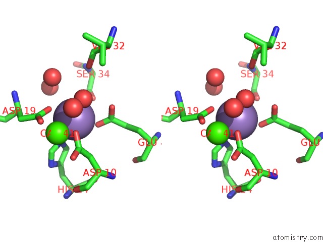
Stereo pair view
Mono view
Stereo pair view
A full contact list of Manganese with other atoms in the Mn binding
site number 1 of Crystal Structure of the Complex of Concanavalin A with A Tripeptide Ypy within 5.0Å range:
|
Reference:
Z.Zhang,
M.Qian,
Q.Huang,
Y.Jia,
Y.Tang,
K.Wang,
D.Cui,
M.Li.
Crystal Structure of the Complex of Concanavalin A and Tripeptide J.Protein Chem. V. 20 59 2001.
ISSN: ISSN 0277-8033
PubMed: 11330349
DOI: 10.1023/A:1011053330536
Page generated: Sat Oct 5 10:54:01 2024
ISSN: ISSN 0277-8033
PubMed: 11330349
DOI: 10.1023/A:1011053330536
Last articles
Zn in 9J0NZn in 9J0O
Zn in 9J0P
Zn in 9FJX
Zn in 9EKB
Zn in 9C0F
Zn in 9CAH
Zn in 9CH0
Zn in 9CH3
Zn in 9CH1