Manganese »
PDB 1ho5-1j53 »
1hqf »
Manganese in PDB 1hqf: Crystal Structure of the Binuclear Manganese Metalloenzyme Arginase Complexed with N-Hydroxy-L-Arginine
Enzymatic activity of Crystal Structure of the Binuclear Manganese Metalloenzyme Arginase Complexed with N-Hydroxy-L-Arginine
All present enzymatic activity of Crystal Structure of the Binuclear Manganese Metalloenzyme Arginase Complexed with N-Hydroxy-L-Arginine:
3.5.3.1;
3.5.3.1;
Protein crystallography data
The structure of Crystal Structure of the Binuclear Manganese Metalloenzyme Arginase Complexed with N-Hydroxy-L-Arginine, PDB code: 1hqf
was solved by
J.D.Cox,
E.Cama,
D.M.Colleluori,
D.E.Ash,
D.W.Christianson,
with X-Ray Crystallography technique. A brief refinement statistics is given in the table below:
| Resolution Low / High (Å) | 30.00 / 2.90 |
| Space group | P 32 |
| Cell size a, b, c (Å), α, β, γ (°) | 88.000, 88.000, 112.000, 90.00, 90.00, 120.00 |
| R / Rfree (%) | 26.5 / 28.9 |
Manganese Binding Sites:
The binding sites of Manganese atom in the Crystal Structure of the Binuclear Manganese Metalloenzyme Arginase Complexed with N-Hydroxy-L-Arginine
(pdb code 1hqf). This binding sites where shown within
5.0 Angstroms radius around Manganese atom.
In total 6 binding sites of Manganese where determined in the Crystal Structure of the Binuclear Manganese Metalloenzyme Arginase Complexed with N-Hydroxy-L-Arginine, PDB code: 1hqf:
Jump to Manganese binding site number: 1; 2; 3; 4; 5; 6;
In total 6 binding sites of Manganese where determined in the Crystal Structure of the Binuclear Manganese Metalloenzyme Arginase Complexed with N-Hydroxy-L-Arginine, PDB code: 1hqf:
Jump to Manganese binding site number: 1; 2; 3; 4; 5; 6;
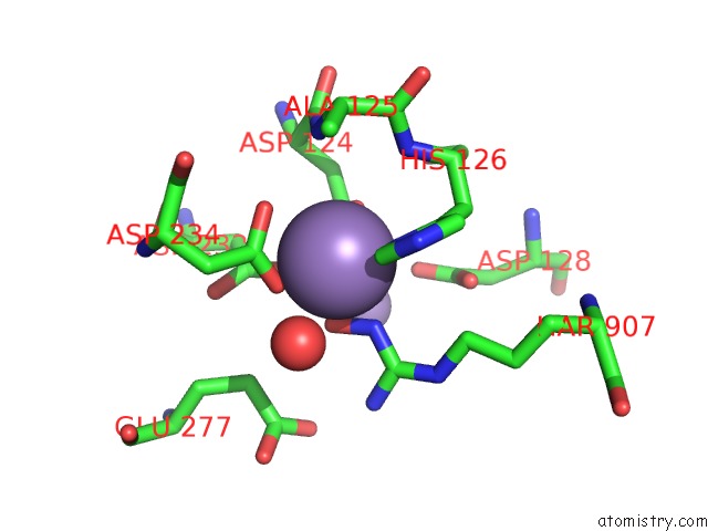
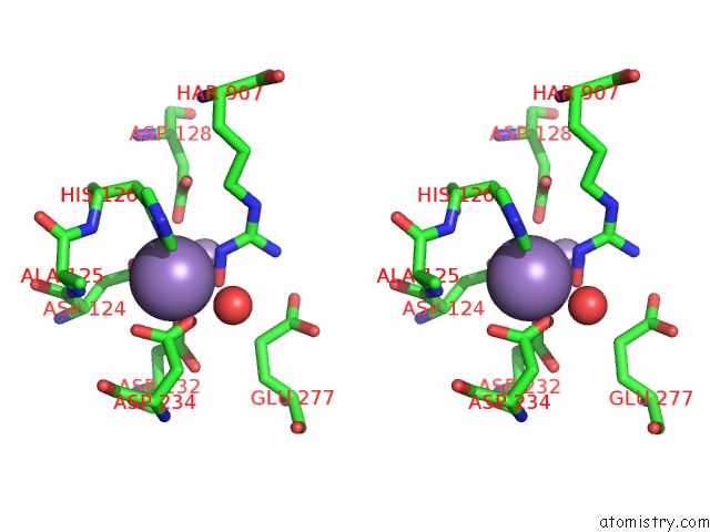
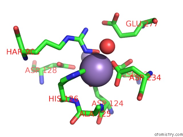
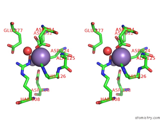
Manganese binding site 1 out of 6 in 1hqf
Go back to
Manganese binding site 1 out
of 6 in the Crystal Structure of the Binuclear Manganese Metalloenzyme Arginase Complexed with N-Hydroxy-L-Arginine
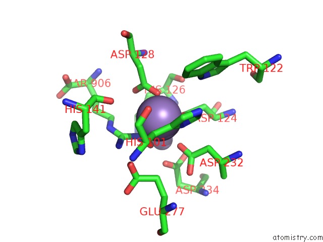
Mono view
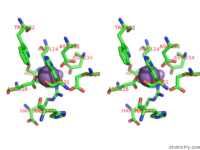
Stereo pair view
Mono view
Stereo pair view
A full contact list of Manganese with other atoms in the Mn binding
site number 1 of Crystal Structure of the Binuclear Manganese Metalloenzyme Arginase Complexed with N-Hydroxy-L-Arginine within 5.0Å range:
|
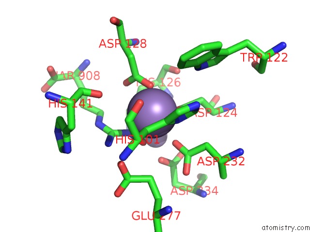
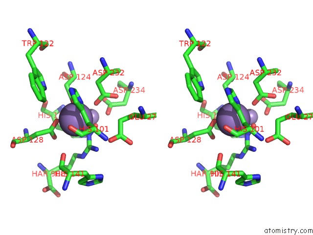
Manganese binding site 2 out of 6 in 1hqf
Go back to
Manganese binding site 2 out
of 6 in the Crystal Structure of the Binuclear Manganese Metalloenzyme Arginase Complexed with N-Hydroxy-L-Arginine
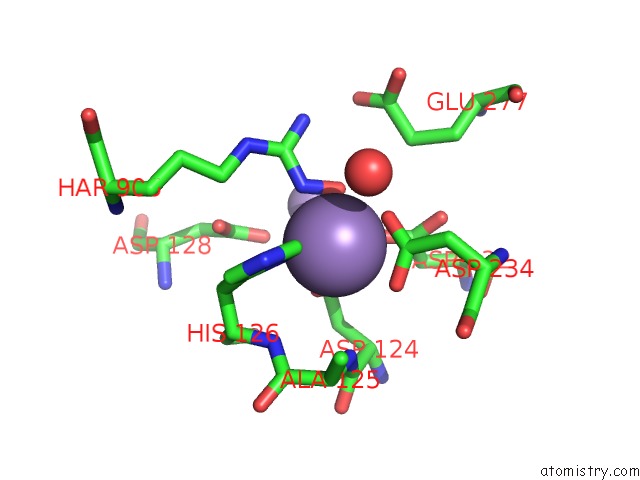
Mono view
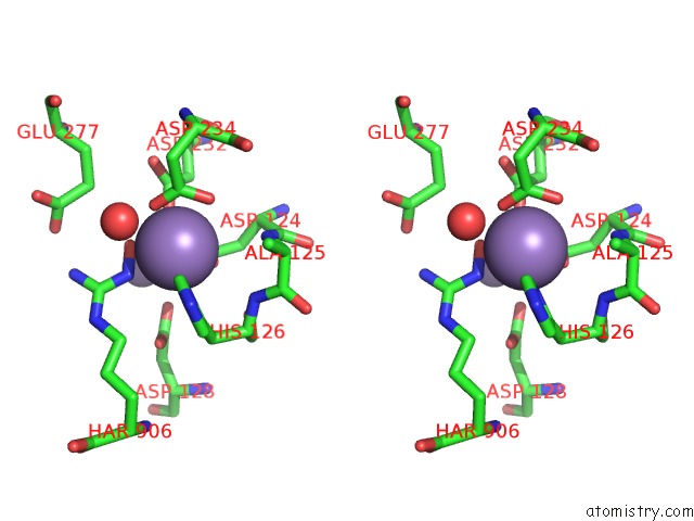
Stereo pair view
Mono view
Stereo pair view
A full contact list of Manganese with other atoms in the Mn binding
site number 2 of Crystal Structure of the Binuclear Manganese Metalloenzyme Arginase Complexed with N-Hydroxy-L-Arginine within 5.0Å range:
|
Manganese binding site 3 out of 6 in 1hqf
Go back to
Manganese binding site 3 out
of 6 in the Crystal Structure of the Binuclear Manganese Metalloenzyme Arginase Complexed with N-Hydroxy-L-Arginine

Mono view

Stereo pair view
Mono view
Stereo pair view
A full contact list of Manganese with other atoms in the Mn binding
site number 3 of Crystal Structure of the Binuclear Manganese Metalloenzyme Arginase Complexed with N-Hydroxy-L-Arginine within 5.0Å range:
|
Manganese binding site 4 out of 6 in 1hqf
Go back to
Manganese binding site 4 out
of 6 in the Crystal Structure of the Binuclear Manganese Metalloenzyme Arginase Complexed with N-Hydroxy-L-Arginine
Mono view
Stereo pair view
Mono view
Stereo pair view
A full contact list of Manganese with other atoms in the Mn binding
site number 4 of Crystal Structure of the Binuclear Manganese Metalloenzyme Arginase Complexed with N-Hydroxy-L-Arginine within 5.0Å range:
|
Manganese binding site 5 out of 6 in 1hqf
Go back to
Manganese binding site 5 out
of 6 in the Crystal Structure of the Binuclear Manganese Metalloenzyme Arginase Complexed with N-Hydroxy-L-Arginine
Mono view
Stereo pair view
Mono view
Stereo pair view
A full contact list of Manganese with other atoms in the Mn binding
site number 5 of Crystal Structure of the Binuclear Manganese Metalloenzyme Arginase Complexed with N-Hydroxy-L-Arginine within 5.0Å range:
|
Manganese binding site 6 out of 6 in 1hqf
Go back to
Manganese binding site 6 out
of 6 in the Crystal Structure of the Binuclear Manganese Metalloenzyme Arginase Complexed with N-Hydroxy-L-Arginine
Mono view
Stereo pair view
Mono view
Stereo pair view
A full contact list of Manganese with other atoms in the Mn binding
site number 6 of Crystal Structure of the Binuclear Manganese Metalloenzyme Arginase Complexed with N-Hydroxy-L-Arginine within 5.0Å range:
|
Reference:
J.D.Cox,
E.Cama,
D.M.Colleluori,
S.Pethe,
J.L.Boucher,
D.Mansuy,
D.E.Ash,
D.W.Christianson.
Mechanistic and Metabolic Inferences From the Binding of Substrate Analogues and Products to Arginase. Biochemistry V. 40 2689 2001.
ISSN: ISSN 0006-2960
PubMed: 11258880
DOI: 10.1021/BI002318+
Page generated: Sat Oct 5 10:54:01 2024
ISSN: ISSN 0006-2960
PubMed: 11258880
DOI: 10.1021/BI002318+
Last articles
Zn in 9MJ5Zn in 9HNW
Zn in 9G0L
Zn in 9FNE
Zn in 9DZN
Zn in 9E0I
Zn in 9D32
Zn in 9DAK
Zn in 8ZXC
Zn in 8ZUF