Manganese »
PDB 1g15-1hkd »
1hkd »
Manganese in PDB 1hkd: Structure of Pea Lectin in Complex with Alpha-Methyl-D-Glucopyranoside
Protein crystallography data
The structure of Structure of Pea Lectin in Complex with Alpha-Methyl-D-Glucopyranoside, PDB code: 1hkd
was solved by
M.B.Shevtsov,
I.N.Tsygannik,
with X-Ray Crystallography technique. A brief refinement statistics is given in the table below:
| Resolution Low / High (Å) | 27.95 / 2.09 |
| Space group | P 21 21 21 |
| Cell size a, b, c (Å), α, β, γ (°) | 50.793, 61.235, 136.005, 90.00, 90.00, 90.00 |
| R / Rfree (%) | 16.3 / 20.9 |
Other elements in 1hkd:
The structure of Structure of Pea Lectin in Complex with Alpha-Methyl-D-Glucopyranoside also contains other interesting chemical elements:
| Calcium | (Ca) | 2 atoms |
Manganese Binding Sites:
The binding sites of Manganese atom in the Structure of Pea Lectin in Complex with Alpha-Methyl-D-Glucopyranoside
(pdb code 1hkd). This binding sites where shown within
5.0 Angstroms radius around Manganese atom.
In total 2 binding sites of Manganese where determined in the Structure of Pea Lectin in Complex with Alpha-Methyl-D-Glucopyranoside, PDB code: 1hkd:
Jump to Manganese binding site number: 1; 2;
In total 2 binding sites of Manganese where determined in the Structure of Pea Lectin in Complex with Alpha-Methyl-D-Glucopyranoside, PDB code: 1hkd:
Jump to Manganese binding site number: 1; 2;
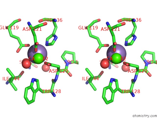
Manganese binding site 1 out of 2 in 1hkd
Go back to
Manganese binding site 1 out
of 2 in the Structure of Pea Lectin in Complex with Alpha-Methyl-D-Glucopyranoside
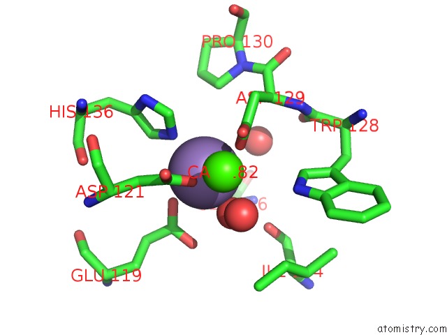
Mono view

Stereo pair view
Mono view
Stereo pair view
A full contact list of Manganese with other atoms in the Mn binding
site number 1 of Structure of Pea Lectin in Complex with Alpha-Methyl-D-Glucopyranoside within 5.0Å range:
|
Manganese binding site 2 out of 2 in 1hkd
Go back to
Manganese binding site 2 out
of 2 in the Structure of Pea Lectin in Complex with Alpha-Methyl-D-Glucopyranoside

Mono view
Stereo pair view
Mono view
Stereo pair view
A full contact list of Manganese with other atoms in the Mn binding
site number 2 of Structure of Pea Lectin in Complex with Alpha-Methyl-D-Glucopyranoside within 5.0Å range:
|
Reference:
M.B.Shevtsov,
I.N.Tsygannik.
Structure of Pea Lectin in Complex with Alpha-Methyl-D-Glucopyranoside To Be Published.
Page generated: Sat Oct 5 10:52:51 2024
Last articles
Ca in 2VZRCa in 2VZQ
Ca in 2VZP
Ca in 2VYP
Ca in 2VYO
Ca in 2VZ1
Ca in 2VY0
Ca in 2VVF
Ca in 2VWO
Ca in 2VWN