Manganese »
PDB 1g15-1hkd »
1gg1 »
Manganese in PDB 1gg1: Crystal Structure Analysis of Dahp Synthase in Complex with MN2+ and 2-Phosphoglycolate
Enzymatic activity of Crystal Structure Analysis of Dahp Synthase in Complex with MN2+ and 2-Phosphoglycolate
All present enzymatic activity of Crystal Structure Analysis of Dahp Synthase in Complex with MN2+ and 2-Phosphoglycolate:
4.1.2.15;
4.1.2.15;
Protein crystallography data
The structure of Crystal Structure Analysis of Dahp Synthase in Complex with MN2+ and 2-Phosphoglycolate, PDB code: 1gg1
was solved by
T.Wagner,
I.A.Shumilin,
R.Bauerle,
R.H.Kretsinger,
with X-Ray Crystallography technique. A brief refinement statistics is given in the table below:
| Resolution Low / High (Å) | 20.00 / 2.00 |
| Space group | C 1 2 1 |
| Cell size a, b, c (Å), α, β, γ (°) | 210.357, 53.188, 149.392, 90.00, 116.09, 90.00 |
| R / Rfree (%) | 20.9 / 23.7 |
Manganese Binding Sites:
The binding sites of Manganese atom in the Crystal Structure Analysis of Dahp Synthase in Complex with MN2+ and 2-Phosphoglycolate
(pdb code 1gg1). This binding sites where shown within
5.0 Angstroms radius around Manganese atom.
In total 4 binding sites of Manganese where determined in the Crystal Structure Analysis of Dahp Synthase in Complex with MN2+ and 2-Phosphoglycolate, PDB code: 1gg1:
Jump to Manganese binding site number: 1; 2; 3; 4;
In total 4 binding sites of Manganese where determined in the Crystal Structure Analysis of Dahp Synthase in Complex with MN2+ and 2-Phosphoglycolate, PDB code: 1gg1:
Jump to Manganese binding site number: 1; 2; 3; 4;
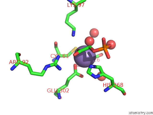
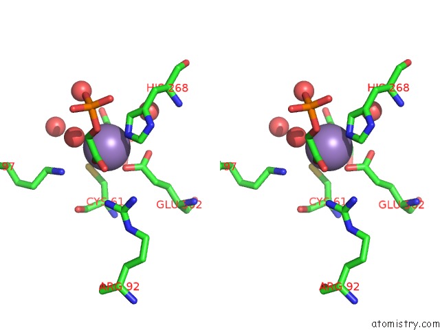
Manganese binding site 1 out of 4 in 1gg1
Go back to
Manganese binding site 1 out
of 4 in the Crystal Structure Analysis of Dahp Synthase in Complex with MN2+ and 2-Phosphoglycolate
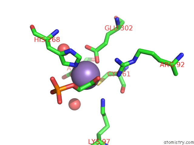
Mono view
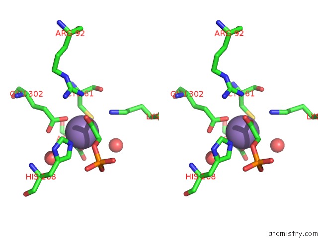
Stereo pair view
Mono view
Stereo pair view
A full contact list of Manganese with other atoms in the Mn binding
site number 1 of Crystal Structure Analysis of Dahp Synthase in Complex with MN2+ and 2-Phosphoglycolate within 5.0Å range:
|
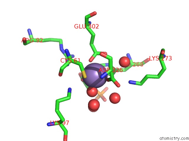
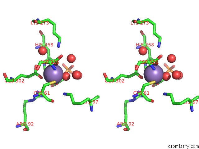
Manganese binding site 2 out of 4 in 1gg1
Go back to
Manganese binding site 2 out
of 4 in the Crystal Structure Analysis of Dahp Synthase in Complex with MN2+ and 2-Phosphoglycolate
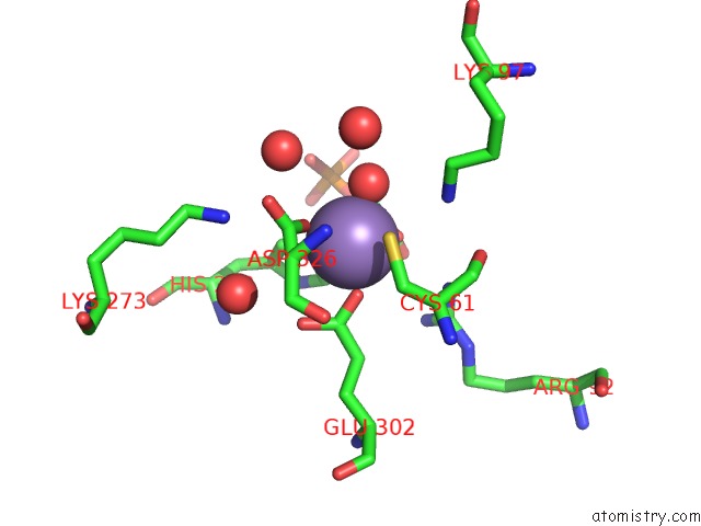
Mono view
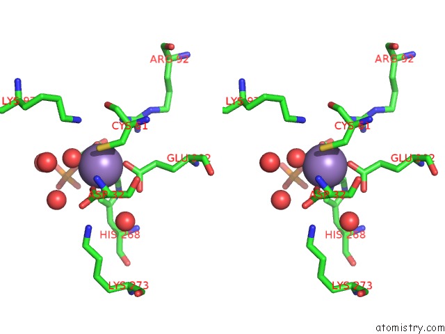
Stereo pair view
Mono view
Stereo pair view
A full contact list of Manganese with other atoms in the Mn binding
site number 2 of Crystal Structure Analysis of Dahp Synthase in Complex with MN2+ and 2-Phosphoglycolate within 5.0Å range:
|
Manganese binding site 3 out of 4 in 1gg1
Go back to
Manganese binding site 3 out
of 4 in the Crystal Structure Analysis of Dahp Synthase in Complex with MN2+ and 2-Phosphoglycolate
Mono view
Stereo pair view
Mono view
Stereo pair view
A full contact list of Manganese with other atoms in the Mn binding
site number 3 of Crystal Structure Analysis of Dahp Synthase in Complex with MN2+ and 2-Phosphoglycolate within 5.0Å range:
|
Manganese binding site 4 out of 4 in 1gg1
Go back to
Manganese binding site 4 out
of 4 in the Crystal Structure Analysis of Dahp Synthase in Complex with MN2+ and 2-Phosphoglycolate
Mono view
Stereo pair view
Mono view
Stereo pair view
A full contact list of Manganese with other atoms in the Mn binding
site number 4 of Crystal Structure Analysis of Dahp Synthase in Complex with MN2+ and 2-Phosphoglycolate within 5.0Å range:
|
Reference:
T.Wagner,
I.A.Shumilin,
R.Bauerle,
R.H.Kretsinger.
Structure of 3-Deoxy-D-Arabino-Heptulosonate-7-Phosphate Synthase From Escherichia Coli: Comparison of the Mn(2+)*2-Phosphoglycolate and the Pb(2+)*2-Phosphoenolpyruvate Complexes and Implications For Catalysis. J.Mol.Biol. V. 301 389 2000.
ISSN: ISSN 0022-2836
PubMed: 10926516
DOI: 10.1006/JMBI.2000.3957
Page generated: Sat Aug 16 07:48:53 2025
ISSN: ISSN 0022-2836
PubMed: 10926516
DOI: 10.1006/JMBI.2000.3957
Last articles
Mn in 5OMVMn in 5OMQ
Mn in 5OEY
Mn in 5OMF
Mn in 5OF3
Mn in 5OGL
Mn in 5O5L
Mn in 5O5K
Mn in 5O7F
Mn in 5O6N