Manganese »
PDB 1g15-1hkd »
1g8w »
Manganese in PDB 1g8w: Improved Structure of Phytohemagglutinin-L From the Kidney Bean
Protein crystallography data
The structure of Improved Structure of Phytohemagglutinin-L From the Kidney Bean, PDB code: 1g8w
was solved by
L.Buts,
T.W.Hamelryck,
M.Dao-Thi,
R.Loris,
L.Wyns,
M.E.Etzler,
with X-Ray Crystallography technique. A brief refinement statistics is given in the table below:
| Resolution Low / High (Å) | 20.00 / 2.80 |
| Space group | C 1 2 1 |
| Cell size a, b, c (Å), α, β, γ (°) | 106.479, 121.278, 89.957, 90.00, 93.18, 90.00 |
| R / Rfree (%) | 18.1 / 21.2 |
Other elements in 1g8w:
The structure of Improved Structure of Phytohemagglutinin-L From the Kidney Bean also contains other interesting chemical elements:
| Calcium | (Ca) | 4 atoms |
Manganese Binding Sites:
The binding sites of Manganese atom in the Improved Structure of Phytohemagglutinin-L From the Kidney Bean
(pdb code 1g8w). This binding sites where shown within
5.0 Angstroms radius around Manganese atom.
In total 4 binding sites of Manganese where determined in the Improved Structure of Phytohemagglutinin-L From the Kidney Bean, PDB code: 1g8w:
Jump to Manganese binding site number: 1; 2; 3; 4;
In total 4 binding sites of Manganese where determined in the Improved Structure of Phytohemagglutinin-L From the Kidney Bean, PDB code: 1g8w:
Jump to Manganese binding site number: 1; 2; 3; 4;
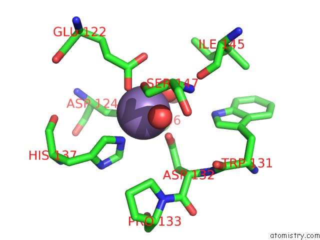
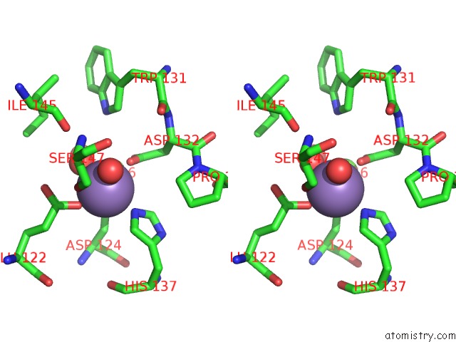
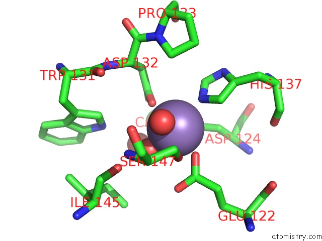
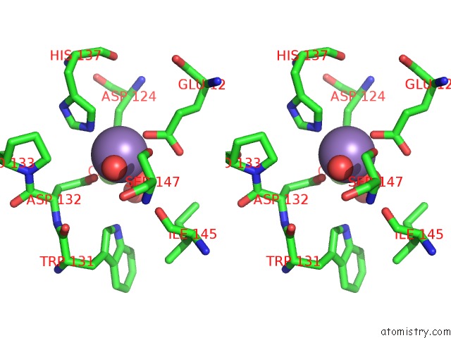
Manganese binding site 1 out of 4 in 1g8w
Go back to
Manganese binding site 1 out
of 4 in the Improved Structure of Phytohemagglutinin-L From the Kidney Bean
Mono view
Stereo pair view
Mono view
Stereo pair view
A full contact list of Manganese with other atoms in the Mn binding
site number 1 of Improved Structure of Phytohemagglutinin-L From the Kidney Bean within 5.0Å range:
|
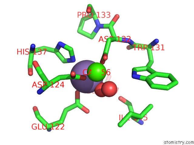
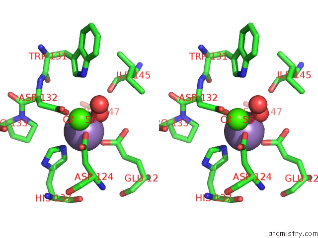
Manganese binding site 2 out of 4 in 1g8w
Go back to
Manganese binding site 2 out
of 4 in the Improved Structure of Phytohemagglutinin-L From the Kidney Bean
Mono view
Stereo pair view
Mono view
Stereo pair view
A full contact list of Manganese with other atoms in the Mn binding
site number 2 of Improved Structure of Phytohemagglutinin-L From the Kidney Bean within 5.0Å range:
|
Manganese binding site 3 out of 4 in 1g8w
Go back to
Manganese binding site 3 out
of 4 in the Improved Structure of Phytohemagglutinin-L From the Kidney Bean

Mono view

Stereo pair view
Mono view
Stereo pair view
A full contact list of Manganese with other atoms in the Mn binding
site number 3 of Improved Structure of Phytohemagglutinin-L From the Kidney Bean within 5.0Å range:
|
Manganese binding site 4 out of 4 in 1g8w
Go back to
Manganese binding site 4 out
of 4 in the Improved Structure of Phytohemagglutinin-L From the Kidney Bean
Mono view
Stereo pair view
Mono view
Stereo pair view
A full contact list of Manganese with other atoms in the Mn binding
site number 4 of Improved Structure of Phytohemagglutinin-L From the Kidney Bean within 5.0Å range:
|
Reference:
L.Buts,
M.H.Dao-Thi,
R.Loris,
L.Wyns,
M.Etzler,
T.Hamelryck.
Weak Protein-Protein Interactions in Lectins: the Crystal Structure of A Vegetative Lectin From the Legume Dolichos Biflorus. J.Mol.Biol. V. 309 193 2001.
ISSN: ISSN 0022-2836
PubMed: 11491289
DOI: 10.1006/JMBI.2001.4639
Page generated: Sat Oct 5 10:31:32 2024
ISSN: ISSN 0022-2836
PubMed: 11491289
DOI: 10.1006/JMBI.2001.4639
Last articles
Fe in 2YXOFe in 2YRS
Fe in 2YXC
Fe in 2YNM
Fe in 2YVJ
Fe in 2YP1
Fe in 2YU2
Fe in 2YU1
Fe in 2YQB
Fe in 2YOO