Manganese »
PDB 1ciw-1en5 »
1els »
Manganese in PDB 1els: Catalytic Metal Ion Binding in Enolase: the Crystal Structure of Enolase-MN2+-Phosphonoacetohydroxamate Complex at 2.4 Angstroms Resolution
Enzymatic activity of Catalytic Metal Ion Binding in Enolase: the Crystal Structure of Enolase-MN2+-Phosphonoacetohydroxamate Complex at 2.4 Angstroms Resolution
All present enzymatic activity of Catalytic Metal Ion Binding in Enolase: the Crystal Structure of Enolase-MN2+-Phosphonoacetohydroxamate Complex at 2.4 Angstroms Resolution:
4.2.1.11;
4.2.1.11;
Protein crystallography data
The structure of Catalytic Metal Ion Binding in Enolase: the Crystal Structure of Enolase-MN2+-Phosphonoacetohydroxamate Complex at 2.4 Angstroms Resolution, PDB code: 1els
was solved by
E.Zhang,
M.Hatada,
J.M.Brewer,
L.Lebioda,
with X-Ray Crystallography technique. A brief refinement statistics is given in the table below:
| Resolution Low / High (Å) | N/A / 2.40 |
| Space group | P 42 21 2 |
| Cell size a, b, c (Å), α, β, γ (°) | 124.100, 124.100, 66.900, 90.00, 90.00, 90.00 |
| R / Rfree (%) | n/a / n/a |
Manganese Binding Sites:
The binding sites of Manganese atom in the Catalytic Metal Ion Binding in Enolase: the Crystal Structure of Enolase-MN2+-Phosphonoacetohydroxamate Complex at 2.4 Angstroms Resolution
(pdb code 1els). This binding sites where shown within
5.0 Angstroms radius around Manganese atom.
In total 2 binding sites of Manganese where determined in the Catalytic Metal Ion Binding in Enolase: the Crystal Structure of Enolase-MN2+-Phosphonoacetohydroxamate Complex at 2.4 Angstroms Resolution, PDB code: 1els:
Jump to Manganese binding site number: 1; 2;
In total 2 binding sites of Manganese where determined in the Catalytic Metal Ion Binding in Enolase: the Crystal Structure of Enolase-MN2+-Phosphonoacetohydroxamate Complex at 2.4 Angstroms Resolution, PDB code: 1els:
Jump to Manganese binding site number: 1; 2;
Manganese binding site 1 out of 2 in 1els
Go back to
Manganese binding site 1 out
of 2 in the Catalytic Metal Ion Binding in Enolase: the Crystal Structure of Enolase-MN2+-Phosphonoacetohydroxamate Complex at 2.4 Angstroms Resolution

Mono view
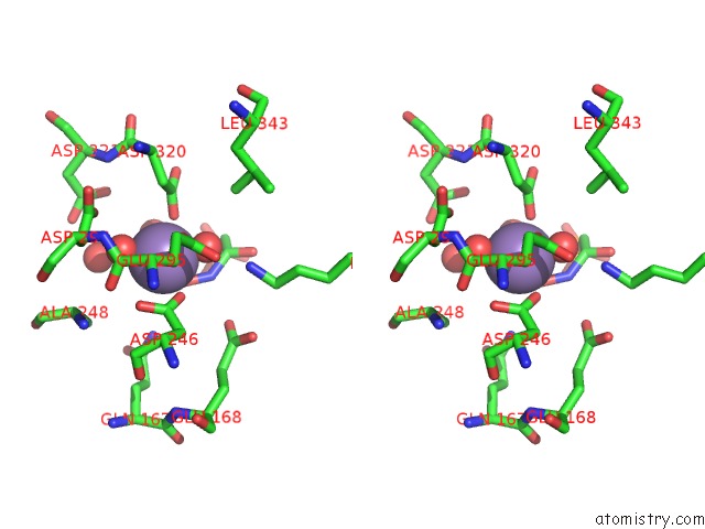
Stereo pair view
Mono view
Stereo pair view
A full contact list of Manganese with other atoms in the Mn binding
site number 1 of Catalytic Metal Ion Binding in Enolase: the Crystal Structure of Enolase-MN2+-Phosphonoacetohydroxamate Complex at 2.4 Angstroms Resolution within 5.0Å range:
|
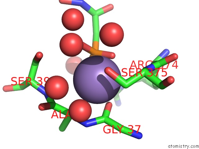
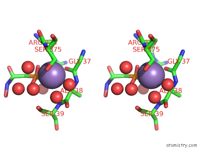
Manganese binding site 2 out of 2 in 1els
Go back to
Manganese binding site 2 out
of 2 in the Catalytic Metal Ion Binding in Enolase: the Crystal Structure of Enolase-MN2+-Phosphonoacetohydroxamate Complex at 2.4 Angstroms Resolution
Mono view
Stereo pair view
Mono view
Stereo pair view
A full contact list of Manganese with other atoms in the Mn binding
site number 2 of Catalytic Metal Ion Binding in Enolase: the Crystal Structure of Enolase-MN2+-Phosphonoacetohydroxamate Complex at 2.4 Angstroms Resolution within 5.0Å range:
|
Reference:
E.Zhang,
M.Hatada,
J.M.Brewer,
L.Lebioda.
Catalytic Metal Ion Binding in Enolase: the Crystal Structure of An Enolase-MN2+-Phosphonoacetohydroxamate Complex at 2.4-A Resolution. Biochemistry V. 33 6295 1994.
ISSN: ISSN 0006-2960
PubMed: 8193144
DOI: 10.1021/BI00186A032
Page generated: Sat Oct 5 10:10:58 2024
ISSN: ISSN 0006-2960
PubMed: 8193144
DOI: 10.1021/BI00186A032
Last articles
Zn in 9J0NZn in 9J0O
Zn in 9J0P
Zn in 9FJX
Zn in 9EKB
Zn in 9C0F
Zn in 9CAH
Zn in 9CH0
Zn in 9CH3
Zn in 9CH1