Manganese »
PDB 1ciw-1en5 »
1ejj »
Manganese in PDB 1ejj: Crystal Structural Analysis of Phosphoglycerate Mutase Cocrystallized with 3-Phosphoglycerate
Enzymatic activity of Crystal Structural Analysis of Phosphoglycerate Mutase Cocrystallized with 3-Phosphoglycerate
All present enzymatic activity of Crystal Structural Analysis of Phosphoglycerate Mutase Cocrystallized with 3-Phosphoglycerate:
5.4.2.1;
5.4.2.1;
Protein crystallography data
The structure of Crystal Structural Analysis of Phosphoglycerate Mutase Cocrystallized with 3-Phosphoglycerate, PDB code: 1ejj
was solved by
M.J.Jedrzejas,
M.Chander,
P.Setlow,
G.Krishnasamy,
with X-Ray Crystallography technique. A brief refinement statistics is given in the table below:
| Resolution Low / High (Å) | 20.00 / 1.90 |
| Space group | C 2 2 21 |
| Cell size a, b, c (Å), α, β, γ (°) | 58.486, 206.293, 125.134, 90.00, 90.00, 90.00 |
| R / Rfree (%) | 20.7 / 24.7 |
Manganese Binding Sites:
The binding sites of Manganese atom in the Crystal Structural Analysis of Phosphoglycerate Mutase Cocrystallized with 3-Phosphoglycerate
(pdb code 1ejj). This binding sites where shown within
5.0 Angstroms radius around Manganese atom.
In total 2 binding sites of Manganese where determined in the Crystal Structural Analysis of Phosphoglycerate Mutase Cocrystallized with 3-Phosphoglycerate, PDB code: 1ejj:
Jump to Manganese binding site number: 1; 2;
In total 2 binding sites of Manganese where determined in the Crystal Structural Analysis of Phosphoglycerate Mutase Cocrystallized with 3-Phosphoglycerate, PDB code: 1ejj:
Jump to Manganese binding site number: 1; 2;
Manganese binding site 1 out of 2 in 1ejj
Go back to
Manganese binding site 1 out
of 2 in the Crystal Structural Analysis of Phosphoglycerate Mutase Cocrystallized with 3-Phosphoglycerate
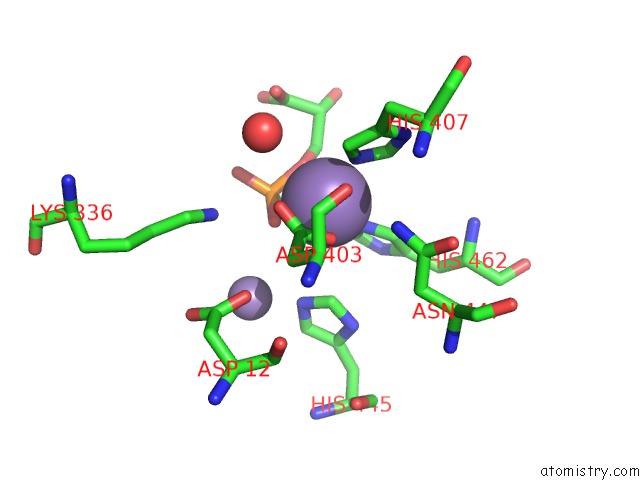
Mono view
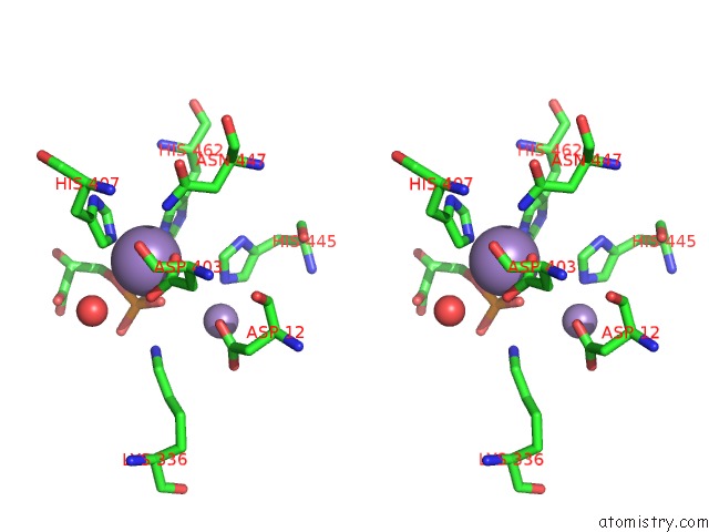
Stereo pair view
Mono view
Stereo pair view
A full contact list of Manganese with other atoms in the Mn binding
site number 1 of Crystal Structural Analysis of Phosphoglycerate Mutase Cocrystallized with 3-Phosphoglycerate within 5.0Å range:
|
Manganese binding site 2 out of 2 in 1ejj
Go back to
Manganese binding site 2 out
of 2 in the Crystal Structural Analysis of Phosphoglycerate Mutase Cocrystallized with 3-Phosphoglycerate
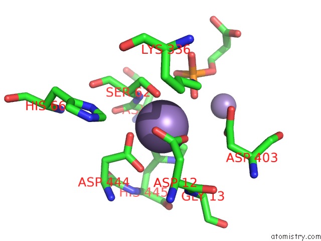
Mono view
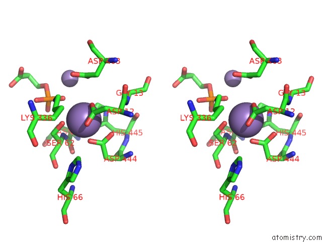
Stereo pair view
Mono view
Stereo pair view
A full contact list of Manganese with other atoms in the Mn binding
site number 2 of Crystal Structural Analysis of Phosphoglycerate Mutase Cocrystallized with 3-Phosphoglycerate within 5.0Å range:
|
Reference:
M.J.Jedrzejas,
M.Chander,
P.Setlow,
G.Krishnasamy.
Structure and Mechanism of Action of A Novel Phosphoglycerate Mutase From Bacillus Stearothermophilus. Embo J. V. 19 1419 2000.
ISSN: ISSN 0261-4189
PubMed: 10747010
DOI: 10.1093/EMBOJ/19.7.1419
Page generated: Sat Oct 5 10:10:39 2024
ISSN: ISSN 0261-4189
PubMed: 10747010
DOI: 10.1093/EMBOJ/19.7.1419
Last articles
Zn in 9J0NZn in 9J0O
Zn in 9J0P
Zn in 9FJX
Zn in 9EKB
Zn in 9C0F
Zn in 9CAH
Zn in 9CH0
Zn in 9CH3
Zn in 9CH1