Manganese »
PDB 1ciw-1en5 »
1dgl »
Manganese in PDB 1dgl: Lectin From Dioclea Grandiflora Complexed to Trimannoside
Protein crystallography data
The structure of Lectin From Dioclea Grandiflora Complexed to Trimannoside, PDB code: 1dgl
was solved by
D.A.Rozwarski,
B.M.Swami,
C.F.Brewer,
J.C.Sacchettini,
with X-Ray Crystallography technique. A brief refinement statistics is given in the table below:
| Resolution Low / High (Å) | 10.00 / 2.40 |
| Space group | P 31 2 1 |
| Cell size a, b, c (Å), α, β, γ (°) | 92.110, 92.110, 202.820, 90.00, 90.00, 120.00 |
| R / Rfree (%) | 18.9 / 25.2 |
Other elements in 1dgl:
The structure of Lectin From Dioclea Grandiflora Complexed to Trimannoside also contains other interesting chemical elements:
| Calcium | (Ca) | 2 atoms |
Manganese Binding Sites:
The binding sites of Manganese atom in the Lectin From Dioclea Grandiflora Complexed to Trimannoside
(pdb code 1dgl). This binding sites where shown within
5.0 Angstroms radius around Manganese atom.
In total 2 binding sites of Manganese where determined in the Lectin From Dioclea Grandiflora Complexed to Trimannoside, PDB code: 1dgl:
Jump to Manganese binding site number: 1; 2;
In total 2 binding sites of Manganese where determined in the Lectin From Dioclea Grandiflora Complexed to Trimannoside, PDB code: 1dgl:
Jump to Manganese binding site number: 1; 2;
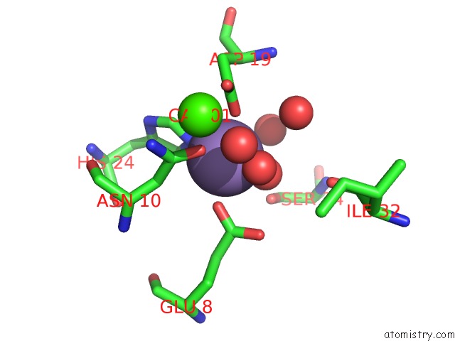
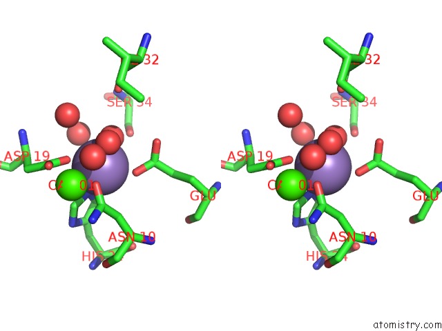
Manganese binding site 1 out of 2 in 1dgl
Go back to
Manganese binding site 1 out
of 2 in the Lectin From Dioclea Grandiflora Complexed to Trimannoside

Mono view

Stereo pair view
Mono view
Stereo pair view
A full contact list of Manganese with other atoms in the Mn binding
site number 1 of Lectin From Dioclea Grandiflora Complexed to Trimannoside within 5.0Å range:
|
Manganese binding site 2 out of 2 in 1dgl
Go back to
Manganese binding site 2 out
of 2 in the Lectin From Dioclea Grandiflora Complexed to Trimannoside
Mono view
Stereo pair view
Mono view
Stereo pair view
A full contact list of Manganese with other atoms in the Mn binding
site number 2 of Lectin From Dioclea Grandiflora Complexed to Trimannoside within 5.0Å range:
|
Reference:
D.A.Rozwarski,
B.M.Swami,
C.F.Brewer,
J.C.Sacchettini.
Crystal Structure of the Lectin From Dioclea Grandiflora Complexed with Core Trimannoside of Asparagine-Linked Carbohydrates. J.Biol.Chem. V. 273 32818 1998.
ISSN: ISSN 0021-9258
PubMed: 9830028
DOI: 10.1074/JBC.273.49.32818
Page generated: Sat Oct 5 10:07:03 2024
ISSN: ISSN 0021-9258
PubMed: 9830028
DOI: 10.1074/JBC.273.49.32818
Last articles
Zn in 9MJ5Zn in 9HNW
Zn in 9G0L
Zn in 9FNE
Zn in 9DZN
Zn in 9E0I
Zn in 9D32
Zn in 9DAK
Zn in 8ZXC
Zn in 8ZUF