Manganese »
PDB 1ciw-1en5 »
1de9 »
Manganese in PDB 1de9: Human APE1 Endonuclease with Bound Abasic Dna and MN2+ Ion
Enzymatic activity of Human APE1 Endonuclease with Bound Abasic Dna and MN2+ Ion
All present enzymatic activity of Human APE1 Endonuclease with Bound Abasic Dna and MN2+ Ion:
4.2.99.18;
4.2.99.18;
Protein crystallography data
The structure of Human APE1 Endonuclease with Bound Abasic Dna and MN2+ Ion, PDB code: 1de9
was solved by
C.D.Mol,
T.Izumi,
S.Mitra,
J.A.Tainer,
with X-Ray Crystallography technique. A brief refinement statistics is given in the table below:
| Resolution Low / High (Å) | 20.00 / 3.00 |
| Space group | P 21 21 21 |
| Cell size a, b, c (Å), α, β, γ (°) | 90.060, 98.350, 101.050, 90.00, 90.00, 90.00 |
| R / Rfree (%) | 18.3 / 27.4 |
Manganese Binding Sites:
The binding sites of Manganese atom in the Human APE1 Endonuclease with Bound Abasic Dna and MN2+ Ion
(pdb code 1de9). This binding sites where shown within
5.0 Angstroms radius around Manganese atom.
In total 2 binding sites of Manganese where determined in the Human APE1 Endonuclease with Bound Abasic Dna and MN2+ Ion, PDB code: 1de9:
Jump to Manganese binding site number: 1; 2;
In total 2 binding sites of Manganese where determined in the Human APE1 Endonuclease with Bound Abasic Dna and MN2+ Ion, PDB code: 1de9:
Jump to Manganese binding site number: 1; 2;
Manganese binding site 1 out of 2 in 1de9
Go back to
Manganese binding site 1 out
of 2 in the Human APE1 Endonuclease with Bound Abasic Dna and MN2+ Ion
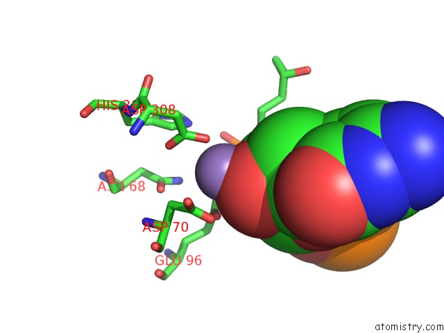
Mono view
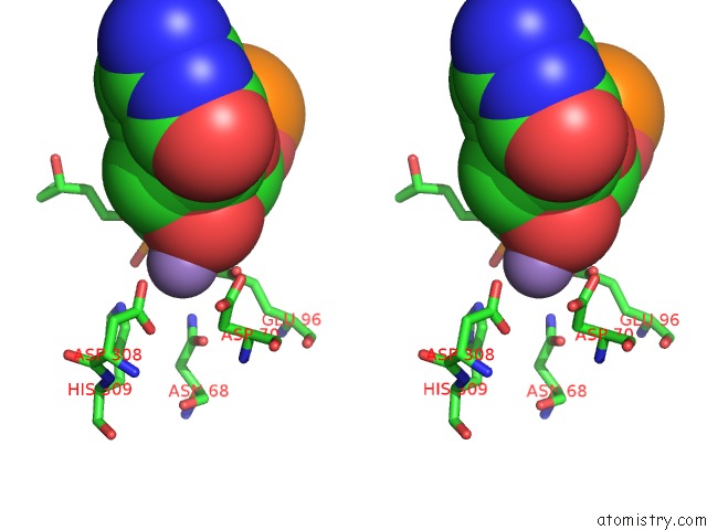
Stereo pair view
Mono view
Stereo pair view
A full contact list of Manganese with other atoms in the Mn binding
site number 1 of Human APE1 Endonuclease with Bound Abasic Dna and MN2+ Ion within 5.0Å range:
|
Manganese binding site 2 out of 2 in 1de9
Go back to
Manganese binding site 2 out
of 2 in the Human APE1 Endonuclease with Bound Abasic Dna and MN2+ Ion
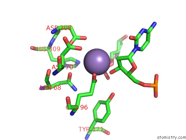
Mono view
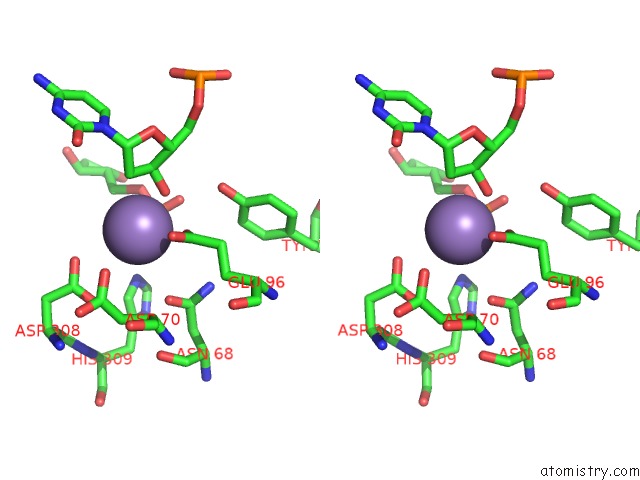
Stereo pair view
Mono view
Stereo pair view
A full contact list of Manganese with other atoms in the Mn binding
site number 2 of Human APE1 Endonuclease with Bound Abasic Dna and MN2+ Ion within 5.0Å range:
|
Reference:
C.D.Mol,
T.Izumi,
S.Mitra,
J.A.Tainer.
Dna-Bound Structures and Mutants Reveal Abasic Dna Binding By APE1 and Dna Repair Coordination Nature V. 403 451 2000.
ISSN: ISSN 0028-0836
PubMed: 10667800
DOI: 10.1038/35000249
Page generated: Sat Oct 5 10:06:33 2024
ISSN: ISSN 0028-0836
PubMed: 10667800
DOI: 10.1038/35000249
Last articles
Zn in 9MJ5Zn in 9HNW
Zn in 9G0L
Zn in 9FNE
Zn in 9DZN
Zn in 9E0I
Zn in 9D32
Zn in 9DAK
Zn in 8ZXC
Zn in 8ZUF