Manganese »
PDB 1ciw-1en5 »
1dck »
Manganese in PDB 1dck: Structure of Unphosphorylated Fixj-N Complexed with MN2+
Protein crystallography data
The structure of Structure of Unphosphorylated Fixj-N Complexed with MN2+, PDB code: 1dck
was solved by
P.Gouet,
B.Fabry,
V.Guillet,
C.Birck,
L.Mourey,
D.Kahn,
J.P.Samama,
with X-Ray Crystallography technique. A brief refinement statistics is given in the table below:
| Resolution Low / High (Å) | 14.45 / 2.00 |
| Space group | P 1 |
| Cell size a, b, c (Å), α, β, γ (°) | 31.700, 42.500, 44.700, 93.20, 103.00, 101.80 |
| R / Rfree (%) | 20.6 / 25.3 |
Manganese Binding Sites:
The binding sites of Manganese atom in the Structure of Unphosphorylated Fixj-N Complexed with MN2+
(pdb code 1dck). This binding sites where shown within
5.0 Angstroms radius around Manganese atom.
In total 3 binding sites of Manganese where determined in the Structure of Unphosphorylated Fixj-N Complexed with MN2+, PDB code: 1dck:
Jump to Manganese binding site number: 1; 2; 3;
In total 3 binding sites of Manganese where determined in the Structure of Unphosphorylated Fixj-N Complexed with MN2+, PDB code: 1dck:
Jump to Manganese binding site number: 1; 2; 3;
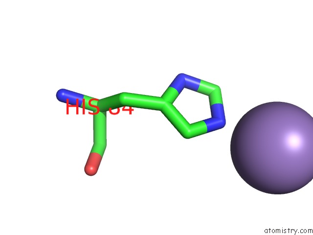
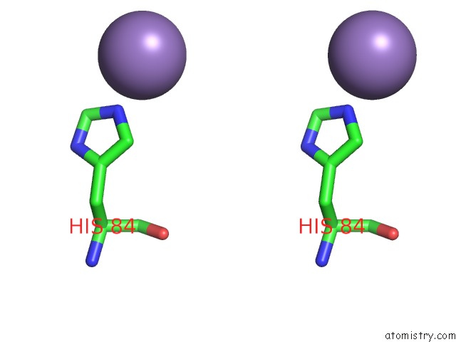
Manganese binding site 1 out of 3 in 1dck
Go back to
Manganese binding site 1 out
of 3 in the Structure of Unphosphorylated Fixj-N Complexed with MN2+
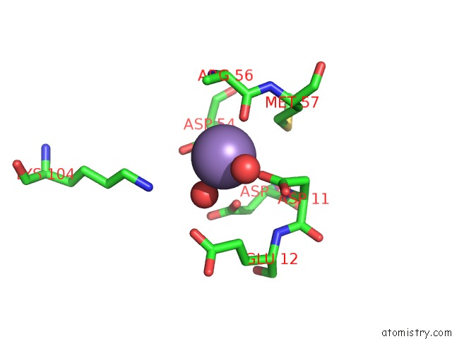
Mono view
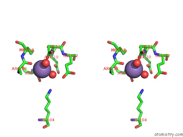
Stereo pair view
Mono view
Stereo pair view
A full contact list of Manganese with other atoms in the Mn binding
site number 1 of Structure of Unphosphorylated Fixj-N Complexed with MN2+ within 5.0Å range:
|
Manganese binding site 2 out of 3 in 1dck
Go back to
Manganese binding site 2 out
of 3 in the Structure of Unphosphorylated Fixj-N Complexed with MN2+
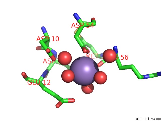
Mono view
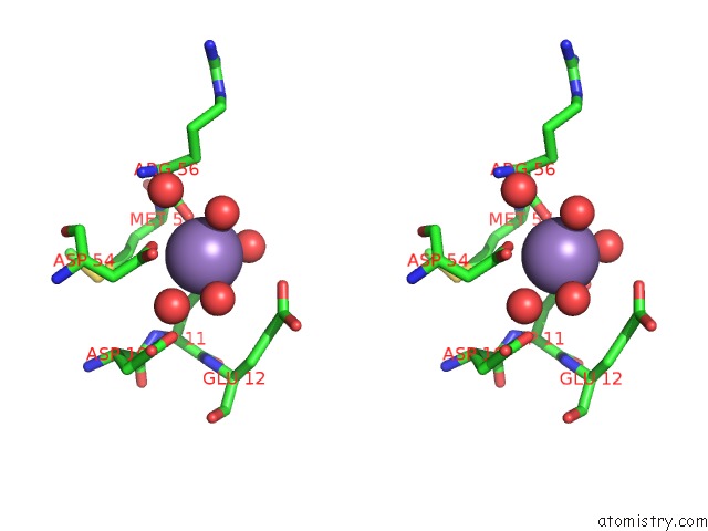
Stereo pair view
Mono view
Stereo pair view
A full contact list of Manganese with other atoms in the Mn binding
site number 2 of Structure of Unphosphorylated Fixj-N Complexed with MN2+ within 5.0Å range:
|
Manganese binding site 3 out of 3 in 1dck
Go back to
Manganese binding site 3 out
of 3 in the Structure of Unphosphorylated Fixj-N Complexed with MN2+
Mono view
Stereo pair view
Mono view
Stereo pair view
A full contact list of Manganese with other atoms in the Mn binding
site number 3 of Structure of Unphosphorylated Fixj-N Complexed with MN2+ within 5.0Å range:
|
Reference:
P.Gouet,
B.Fabry,
V.Guillet,
C.Birck,
L.Mourey,
D.Kahn,
J.P.Samama.
Structural Transitions in the Fixj Receiver Domain. Structure Fold.Des. V. 7 1517 1999.
ISSN: ISSN 0969-2126
PubMed: 10647182
DOI: 10.1016/S0969-2126(00)88342-2
Page generated: Sat Oct 5 10:05:32 2024
ISSN: ISSN 0969-2126
PubMed: 10647182
DOI: 10.1016/S0969-2126(00)88342-2
Last articles
Fe in 2YXOFe in 2YRS
Fe in 2YXC
Fe in 2YNM
Fe in 2YVJ
Fe in 2YP1
Fe in 2YU2
Fe in 2YU1
Fe in 2YQB
Fe in 2YOO