Manganese »
PDB 1ciw-1en5 »
1d5n »
Manganese in PDB 1d5n: Crystal Structure of E. Coli Mnsod at 100K
Enzymatic activity of Crystal Structure of E. Coli Mnsod at 100K
All present enzymatic activity of Crystal Structure of E. Coli Mnsod at 100K:
1.15.1.1;
1.15.1.1;
Protein crystallography data
The structure of Crystal Structure of E. Coli Mnsod at 100K, PDB code: 1d5n
was solved by
G.E.O.Borgstahl,
M.Pokross,
R.Chehab,
A.Sekher,
E.H.Snell,
with X-Ray Crystallography technique. A brief refinement statistics is given in the table below:
| Resolution Low / High (Å) | 20.00 / 1.55 |
| Space group | C 2 2 21 |
| Cell size a, b, c (Å), α, β, γ (°) | 99.110, 107.300, 179.110, 90.00, 90.00, 90.00 |
| R / Rfree (%) | 19.4 / 23.3 |
Manganese Binding Sites:
The binding sites of Manganese atom in the Crystal Structure of E. Coli Mnsod at 100K
(pdb code 1d5n). This binding sites where shown within
5.0 Angstroms radius around Manganese atom.
In total 4 binding sites of Manganese where determined in the Crystal Structure of E. Coli Mnsod at 100K, PDB code: 1d5n:
Jump to Manganese binding site number: 1; 2; 3; 4;
In total 4 binding sites of Manganese where determined in the Crystal Structure of E. Coli Mnsod at 100K, PDB code: 1d5n:
Jump to Manganese binding site number: 1; 2; 3; 4;
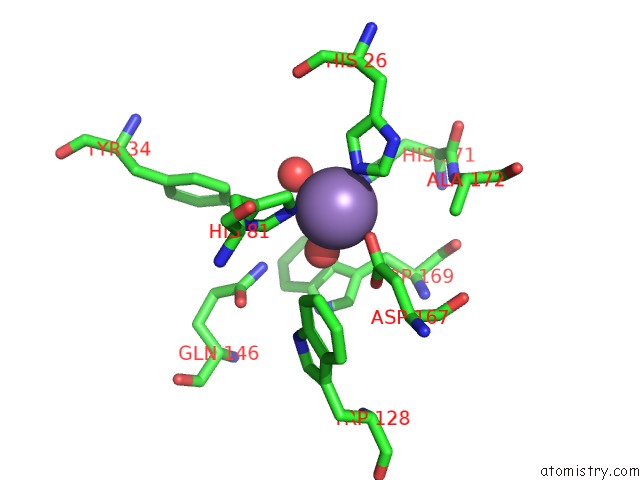
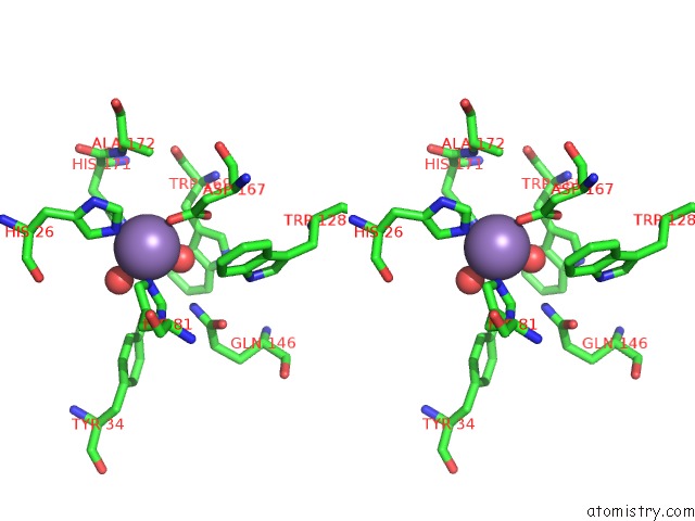
Manganese binding site 1 out of 4 in 1d5n
Go back to
Manganese binding site 1 out
of 4 in the Crystal Structure of E. Coli Mnsod at 100K
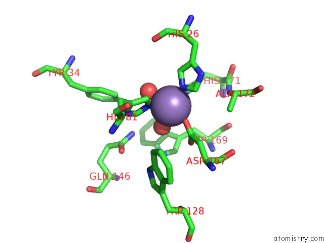
Mono view
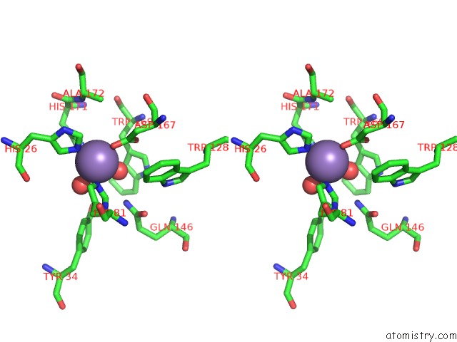
Stereo pair view
Mono view
Stereo pair view
A full contact list of Manganese with other atoms in the Mn binding
site number 1 of Crystal Structure of E. Coli Mnsod at 100K within 5.0Å range:
|
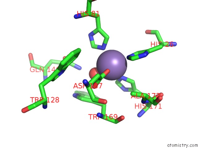
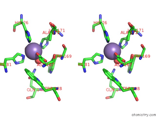
Manganese binding site 2 out of 4 in 1d5n
Go back to
Manganese binding site 2 out
of 4 in the Crystal Structure of E. Coli Mnsod at 100K
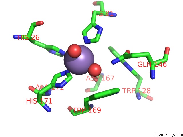
Mono view
Stereo pair view
Mono view
Stereo pair view
A full contact list of Manganese with other atoms in the Mn binding
site number 2 of Crystal Structure of E. Coli Mnsod at 100K within 5.0Å range:
|
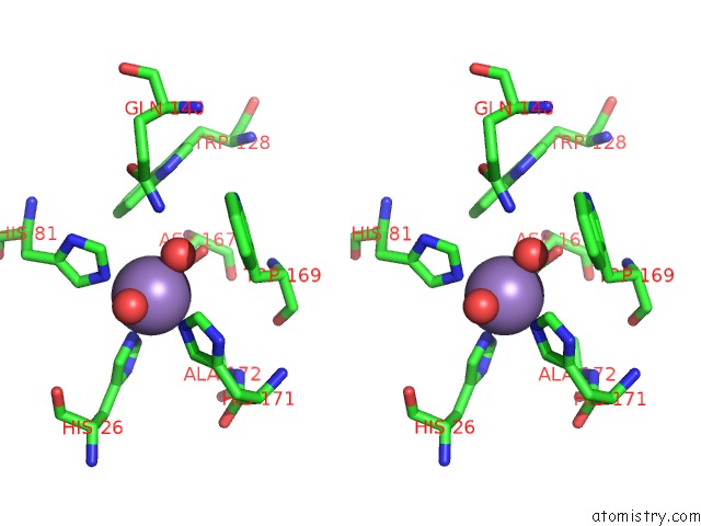
Manganese binding site 3 out of 4 in 1d5n
Go back to
Manganese binding site 3 out
of 4 in the Crystal Structure of E. Coli Mnsod at 100K
Mono view
Stereo pair view
Mono view
Stereo pair view
A full contact list of Manganese with other atoms in the Mn binding
site number 3 of Crystal Structure of E. Coli Mnsod at 100K within 5.0Å range:
|
Manganese binding site 4 out of 4 in 1d5n
Go back to
Manganese binding site 4 out
of 4 in the Crystal Structure of E. Coli Mnsod at 100K
Mono view
Stereo pair view
Mono view
Stereo pair view
A full contact list of Manganese with other atoms in the Mn binding
site number 4 of Crystal Structure of E. Coli Mnsod at 100K within 5.0Å range:
|
Reference:
G.E.Borgstahl,
M.Pokross,
R.Chehab,
A.Sekher,
E.H.Snell.
Cryo-Trapping the Six-Coordinate, Distorted-Octahedral Active Site of Manganese Superoxide Dismutase. J.Mol.Biol. V. 296 951 2000.
ISSN: ISSN 0022-2836
PubMed: 10686094
DOI: 10.1006/JMBI.1999.3506
Page generated: Sat Oct 5 10:04:30 2024
ISSN: ISSN 0022-2836
PubMed: 10686094
DOI: 10.1006/JMBI.1999.3506
Last articles
F in 7KOPF in 7KO0
F in 7KNY
F in 7KM8
F in 7KM9
F in 7KNR
F in 7KKO
F in 7KM7
F in 7KK3
F in 7KLE