Manganese »
PDB 1ciw-1en5 »
1cw4 »
Manganese in PDB 1cw4: Crystal Structure of K230M Isocitrate Dehydrogenase in Complex with Alpha-Ketoglutarate
Enzymatic activity of Crystal Structure of K230M Isocitrate Dehydrogenase in Complex with Alpha-Ketoglutarate
All present enzymatic activity of Crystal Structure of K230M Isocitrate Dehydrogenase in Complex with Alpha-Ketoglutarate:
1.1.1.42;
1.1.1.42;
Protein crystallography data
The structure of Crystal Structure of K230M Isocitrate Dehydrogenase in Complex with Alpha-Ketoglutarate, PDB code: 1cw4
was solved by
M.R.Stroud,
J.Finer-Moore,
with X-Ray Crystallography technique. A brief refinement statistics is given in the table below:
| Resolution Low / High (Å) | 5.00 / 2.10 |
| Space group | P 43 21 2 |
| Cell size a, b, c (Å), α, β, γ (°) | 102.800, 102.800, 150.100, 90.00, 90.00, 90.00 |
| R / Rfree (%) | 18.4 / 22.6 |
Manganese Binding Sites:
The binding sites of Manganese atom in the Crystal Structure of K230M Isocitrate Dehydrogenase in Complex with Alpha-Ketoglutarate
(pdb code 1cw4). This binding sites where shown within
5.0 Angstroms radius around Manganese atom.
In total only one binding site of Manganese was determined in the Crystal Structure of K230M Isocitrate Dehydrogenase in Complex with Alpha-Ketoglutarate, PDB code: 1cw4:
In total only one binding site of Manganese was determined in the Crystal Structure of K230M Isocitrate Dehydrogenase in Complex with Alpha-Ketoglutarate, PDB code: 1cw4:
Manganese binding site 1 out of 1 in 1cw4
Go back to
Manganese binding site 1 out
of 1 in the Crystal Structure of K230M Isocitrate Dehydrogenase in Complex with Alpha-Ketoglutarate
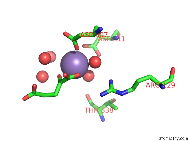
Mono view
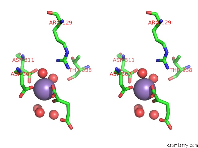
Stereo pair view
Mono view
Stereo pair view
A full contact list of Manganese with other atoms in the Mn binding
site number 1 of Crystal Structure of K230M Isocitrate Dehydrogenase in Complex with Alpha-Ketoglutarate within 5.0Å range:
|
Reference:
D.B.Cherbavaz,
M.E.Lee,
R.M.Stroud,
D.E.Koshland Jr..
Active Site Water Molecules Revealed in the 2.1 A Resolution Structure of A Site-Directed Mutant of Isocitrate Dehydrogenase. J.Mol.Biol. V. 295 377 2000.
ISSN: ISSN 0022-2836
PubMed: 10623532
DOI: 10.1006/JMBI.1999.3195
Page generated: Sat Oct 5 10:03:03 2024
ISSN: ISSN 0022-2836
PubMed: 10623532
DOI: 10.1006/JMBI.1999.3195
Last articles
Zn in 9MJ5Zn in 9HNW
Zn in 9G0L
Zn in 9FNE
Zn in 9DZN
Zn in 9E0I
Zn in 9D32
Zn in 9DAK
Zn in 8ZXC
Zn in 8ZUF