Manganese »
PDB 1ciw-1en5 »
1cq9 »
Manganese in PDB 1cq9: Peanut Lectin-Triclinic Form
Protein crystallography data
The structure of Peanut Lectin-Triclinic Form, PDB code: 1cq9
was solved by
R.Ravishankar,
K.Suguna,
A.Surolia,
M.Vijayan,
with X-Ray Crystallography technique. A brief refinement statistics is given in the table below:
| Resolution Low / High (Å) | 10.00 / 3.50 |
| Space group | P 1 |
| Cell size a, b, c (Å), α, β, γ (°) | 53.640, 71.790, 86.420, 65.35, 77.66, 72.31 |
| R / Rfree (%) | 21.7 / 27.8 |
Other elements in 1cq9:
The structure of Peanut Lectin-Triclinic Form also contains other interesting chemical elements:
| Calcium | (Ca) | 4 atoms |
Manganese Binding Sites:
The binding sites of Manganese atom in the Peanut Lectin-Triclinic Form
(pdb code 1cq9). This binding sites where shown within
5.0 Angstroms radius around Manganese atom.
In total 4 binding sites of Manganese where determined in the Peanut Lectin-Triclinic Form, PDB code: 1cq9:
Jump to Manganese binding site number: 1; 2; 3; 4;
In total 4 binding sites of Manganese where determined in the Peanut Lectin-Triclinic Form, PDB code: 1cq9:
Jump to Manganese binding site number: 1; 2; 3; 4;
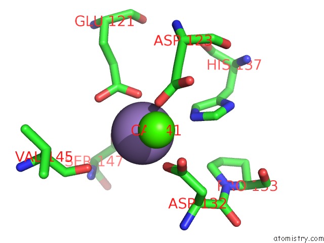
Manganese binding site 1 out of 4 in 1cq9
Go back to
Manganese binding site 1 out
of 4 in the Peanut Lectin-Triclinic Form
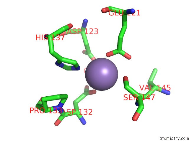
Mono view
Stereo pair view
Mono view
Stereo pair view
A full contact list of Manganese with other atoms in the Mn binding
site number 1 of Peanut Lectin-Triclinic Form within 5.0Å range:
|
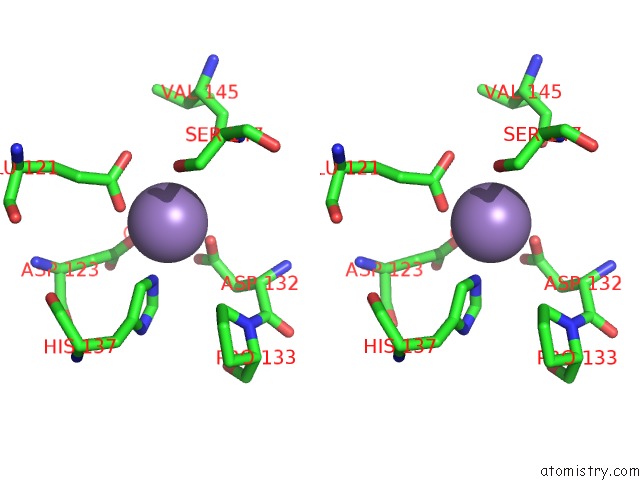
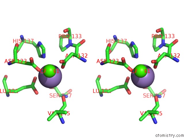
Manganese binding site 2 out of 4 in 1cq9
Go back to
Manganese binding site 2 out
of 4 in the Peanut Lectin-Triclinic Form
Mono view
Stereo pair view
Mono view
Stereo pair view
A full contact list of Manganese with other atoms in the Mn binding
site number 2 of Peanut Lectin-Triclinic Form within 5.0Å range:
|
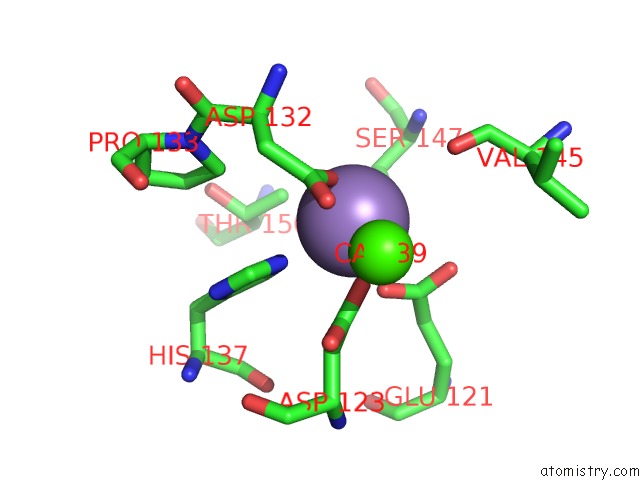
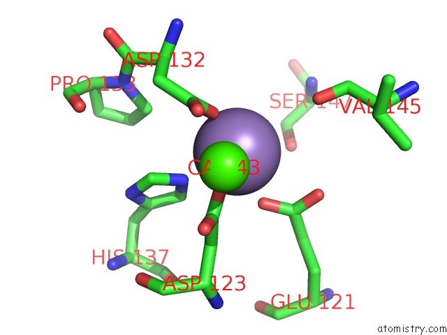
Manganese binding site 3 out of 4 in 1cq9
Go back to
Manganese binding site 3 out
of 4 in the Peanut Lectin-Triclinic Form
Mono view
Stereo pair view
Mono view
Stereo pair view
A full contact list of Manganese with other atoms in the Mn binding
site number 3 of Peanut Lectin-Triclinic Form within 5.0Å range:
|
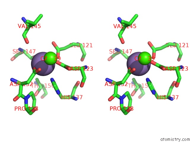
Manganese binding site 4 out of 4 in 1cq9
Go back to
Manganese binding site 4 out
of 4 in the Peanut Lectin-Triclinic Form
Mono view
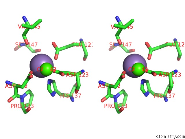
Stereo pair view
Mono view
Stereo pair view
A full contact list of Manganese with other atoms in the Mn binding
site number 4 of Peanut Lectin-Triclinic Form within 5.0Å range:
|
Reference:
R.Ravishankar,
C.J.Thomas,
K.Suguna,
A.Surolia,
M.Vijayan.
Crystal Structures of the Peanut Lectin-Lactose Complex at Acidic pH: Retention of Unusual Quaternary Structure, Empty and Carbohydrate Bound Combining Sites, Molecular Mimicry and Crystal Packing Directed By Interactions at the Combining Site. Proteins V. 43 260 2001.
ISSN: ISSN 0887-3585
PubMed: 11288176
DOI: 10.1002/PROT.1037
Page generated: Sat Oct 5 10:01:45 2024
ISSN: ISSN 0887-3585
PubMed: 11288176
DOI: 10.1002/PROT.1037
Last articles
Zn in 9J0NZn in 9J0O
Zn in 9J0P
Zn in 9FJX
Zn in 9EKB
Zn in 9C0F
Zn in 9CAH
Zn in 9CH0
Zn in 9CH3
Zn in 9CH1