Manganese »
PDB 117e-1cev »
1c39 »
Manganese in PDB 1c39: Structure of Cation-Dependent Mannose 6-Phosphate Receptor Bound to Pentamannosyl Phosphate
Protein crystallography data
The structure of Structure of Cation-Dependent Mannose 6-Phosphate Receptor Bound to Pentamannosyl Phosphate, PDB code: 1c39
was solved by
L.J.Olson,
J.Zhang,
Y.C.Lee,
N.M.Dahms,
J.J.-P.Kim,
with X-Ray Crystallography technique. A brief refinement statistics is given in the table below:
| Resolution Low / High (Å) | 40.00 / 1.85 |
| Space group | P 1 21 1 |
| Cell size a, b, c (Å), α, β, γ (°) | 42.840, 79.310, 55.570, 90.00, 100.33, 90.00 |
| R / Rfree (%) | 21 / 24.6 |
Manganese Binding Sites:
The binding sites of Manganese atom in the Structure of Cation-Dependent Mannose 6-Phosphate Receptor Bound to Pentamannosyl Phosphate
(pdb code 1c39). This binding sites where shown within
5.0 Angstroms radius around Manganese atom.
In total 2 binding sites of Manganese where determined in the Structure of Cation-Dependent Mannose 6-Phosphate Receptor Bound to Pentamannosyl Phosphate, PDB code: 1c39:
Jump to Manganese binding site number: 1; 2;
In total 2 binding sites of Manganese where determined in the Structure of Cation-Dependent Mannose 6-Phosphate Receptor Bound to Pentamannosyl Phosphate, PDB code: 1c39:
Jump to Manganese binding site number: 1; 2;
Manganese binding site 1 out of 2 in 1c39
Go back to
Manganese binding site 1 out
of 2 in the Structure of Cation-Dependent Mannose 6-Phosphate Receptor Bound to Pentamannosyl Phosphate
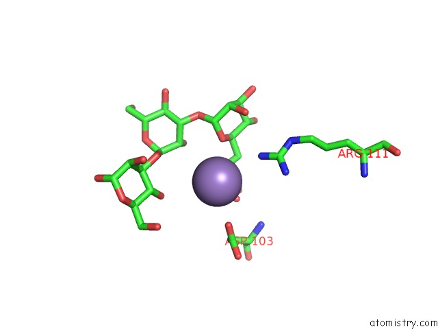
Mono view
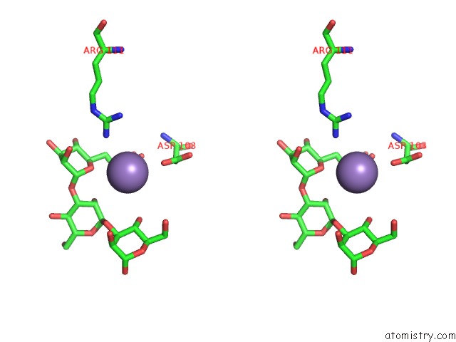
Stereo pair view
Mono view
Stereo pair view
A full contact list of Manganese with other atoms in the Mn binding
site number 1 of Structure of Cation-Dependent Mannose 6-Phosphate Receptor Bound to Pentamannosyl Phosphate within 5.0Å range:
|
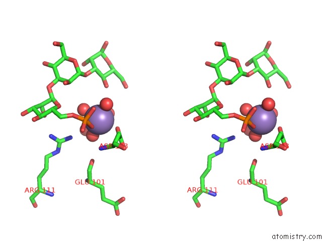
Manganese binding site 2 out of 2 in 1c39
Go back to
Manganese binding site 2 out
of 2 in the Structure of Cation-Dependent Mannose 6-Phosphate Receptor Bound to Pentamannosyl Phosphate
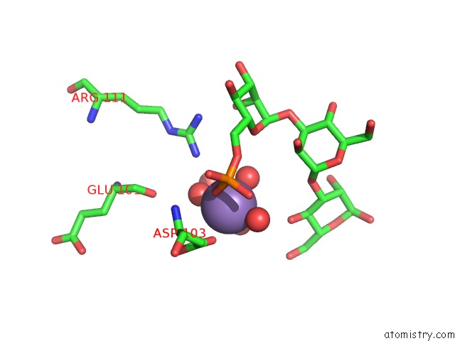
Mono view
Stereo pair view
Mono view
Stereo pair view
A full contact list of Manganese with other atoms in the Mn binding
site number 2 of Structure of Cation-Dependent Mannose 6-Phosphate Receptor Bound to Pentamannosyl Phosphate within 5.0Å range:
|
Reference:
L.J.Olson,
J.Zhang,
Y.C.Lee,
N.M.Dahms,
J.J.Kim.
Structural Basis For Recognition of Phosphorylated High Mannose Oligosaccharides By the Cation-Dependent Mannose 6-Phosphate Receptor. J.Biol.Chem. V. 274 29889 1999.
ISSN: ISSN 0021-9258
PubMed: 10514470
DOI: 10.1074/JBC.274.42.29889
Page generated: Sat Oct 5 09:50:59 2024
ISSN: ISSN 0021-9258
PubMed: 10514470
DOI: 10.1074/JBC.274.42.29889
Last articles
Zn in 9J0NZn in 9J0O
Zn in 9J0P
Zn in 9FJX
Zn in 9EKB
Zn in 9C0F
Zn in 9CAH
Zn in 9CH0
Zn in 9CH3
Zn in 9CH1