Manganese in PDB 8q1x: Structural Analysis of PLD3 Reveals Insights Into the Mechanism of Lysosomal 5' Exonuclease-Mediated Nucleic Acid Degradation
Protein crystallography data
The structure of Structural Analysis of PLD3 Reveals Insights Into the Mechanism of Lysosomal 5' Exonuclease-Mediated Nucleic Acid Degradation, PDB code: 8q1x
was solved by
Y.Roske,
O.Daumke,
M.Damme,
with X-Ray Crystallography technique. A brief refinement statistics is given in the table below:
| Resolution Low / High (Å) | 35.78 / 1.85 |
| Space group | P 1 21 1 |
| Cell size a, b, c (Å), α, β, γ (°) | 60.535, 115.452, 101.335, 90, 106.62, 90 |
| R / Rfree (%) | 17.4 / 19.9 |
Other elements in 8q1x:
The structure of Structural Analysis of PLD3 Reveals Insights Into the Mechanism of Lysosomal 5' Exonuclease-Mediated Nucleic Acid Degradation also contains other interesting chemical elements:
| Magnesium | (Mg) | 2 atoms |
Manganese Binding Sites:
The binding sites of Manganese atom in the Structural Analysis of PLD3 Reveals Insights Into the Mechanism of Lysosomal 5' Exonuclease-Mediated Nucleic Acid Degradation
(pdb code 8q1x). This binding sites where shown within
5.0 Angstroms radius around Manganese atom.
In total 2 binding sites of Manganese where determined in the Structural Analysis of PLD3 Reveals Insights Into the Mechanism of Lysosomal 5' Exonuclease-Mediated Nucleic Acid Degradation, PDB code: 8q1x:
Jump to Manganese binding site number: 1; 2;
In total 2 binding sites of Manganese where determined in the Structural Analysis of PLD3 Reveals Insights Into the Mechanism of Lysosomal 5' Exonuclease-Mediated Nucleic Acid Degradation, PDB code: 8q1x:
Jump to Manganese binding site number: 1; 2;
Manganese binding site 1 out of 2 in 8q1x
Go back to
Manganese binding site 1 out
of 2 in the Structural Analysis of PLD3 Reveals Insights Into the Mechanism of Lysosomal 5' Exonuclease-Mediated Nucleic Acid Degradation
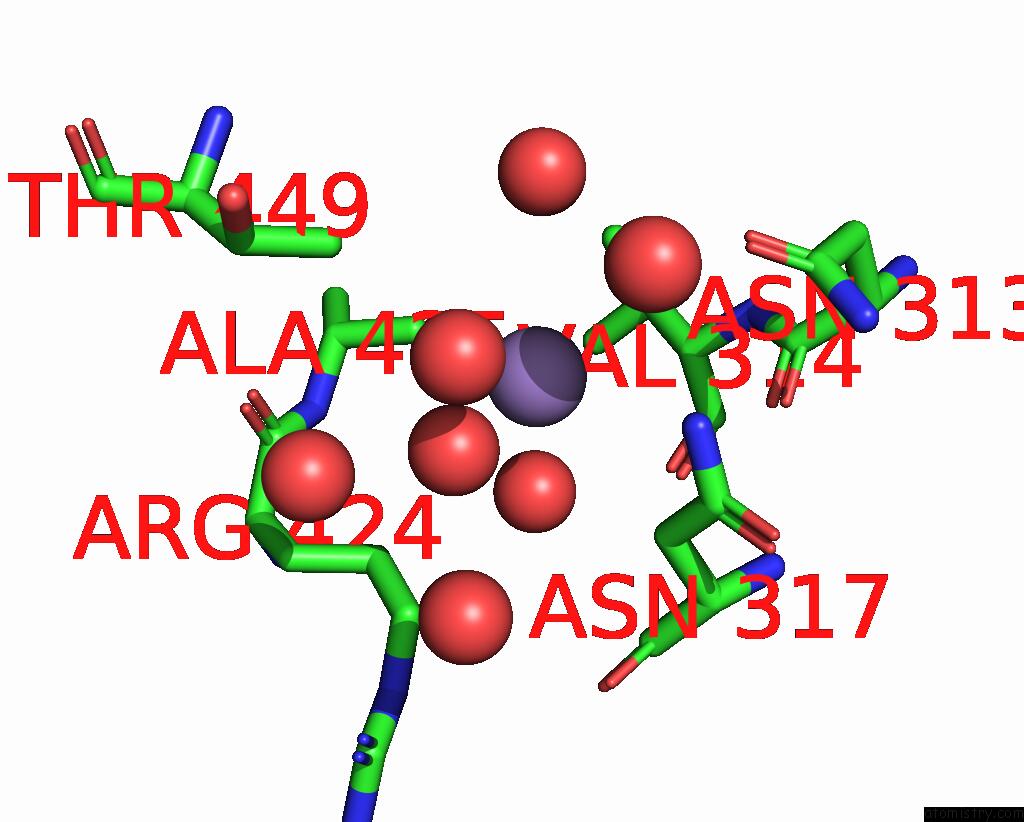
Mono view
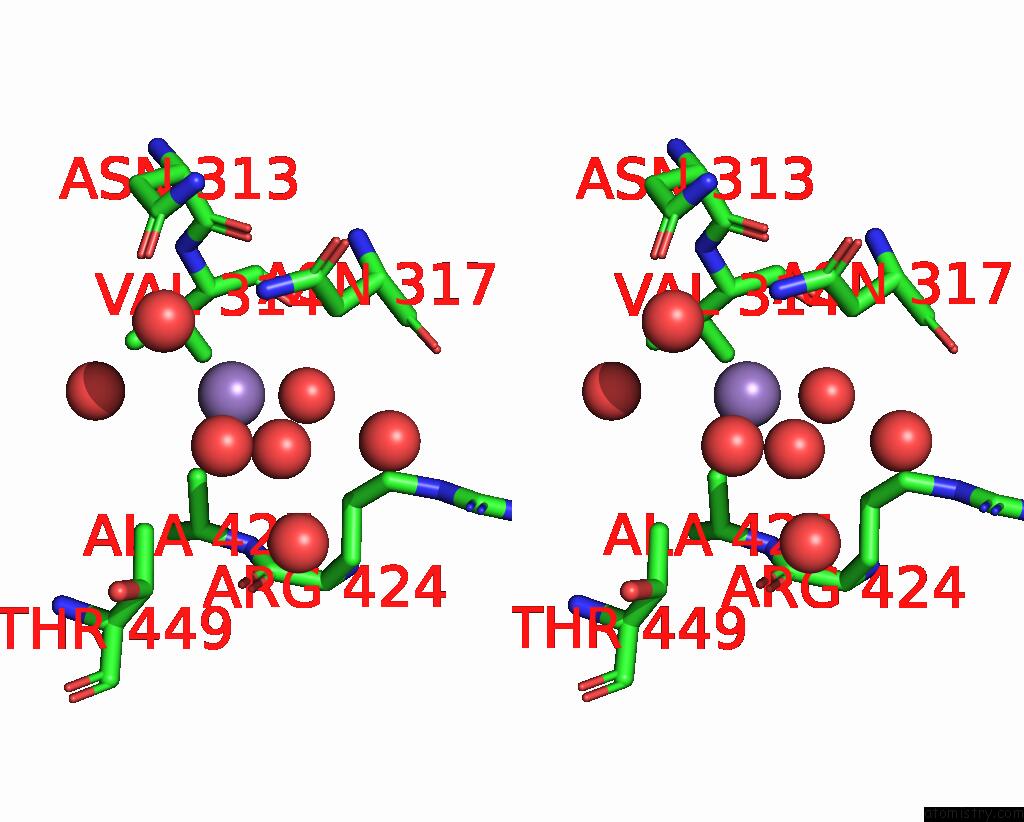
Stereo pair view
Mono view
Stereo pair view
A full contact list of Manganese with other atoms in the Mn binding
site number 1 of Structural Analysis of PLD3 Reveals Insights Into the Mechanism of Lysosomal 5' Exonuclease-Mediated Nucleic Acid Degradation within 5.0Å range:
|
Manganese binding site 2 out of 2 in 8q1x
Go back to
Manganese binding site 2 out
of 2 in the Structural Analysis of PLD3 Reveals Insights Into the Mechanism of Lysosomal 5' Exonuclease-Mediated Nucleic Acid Degradation
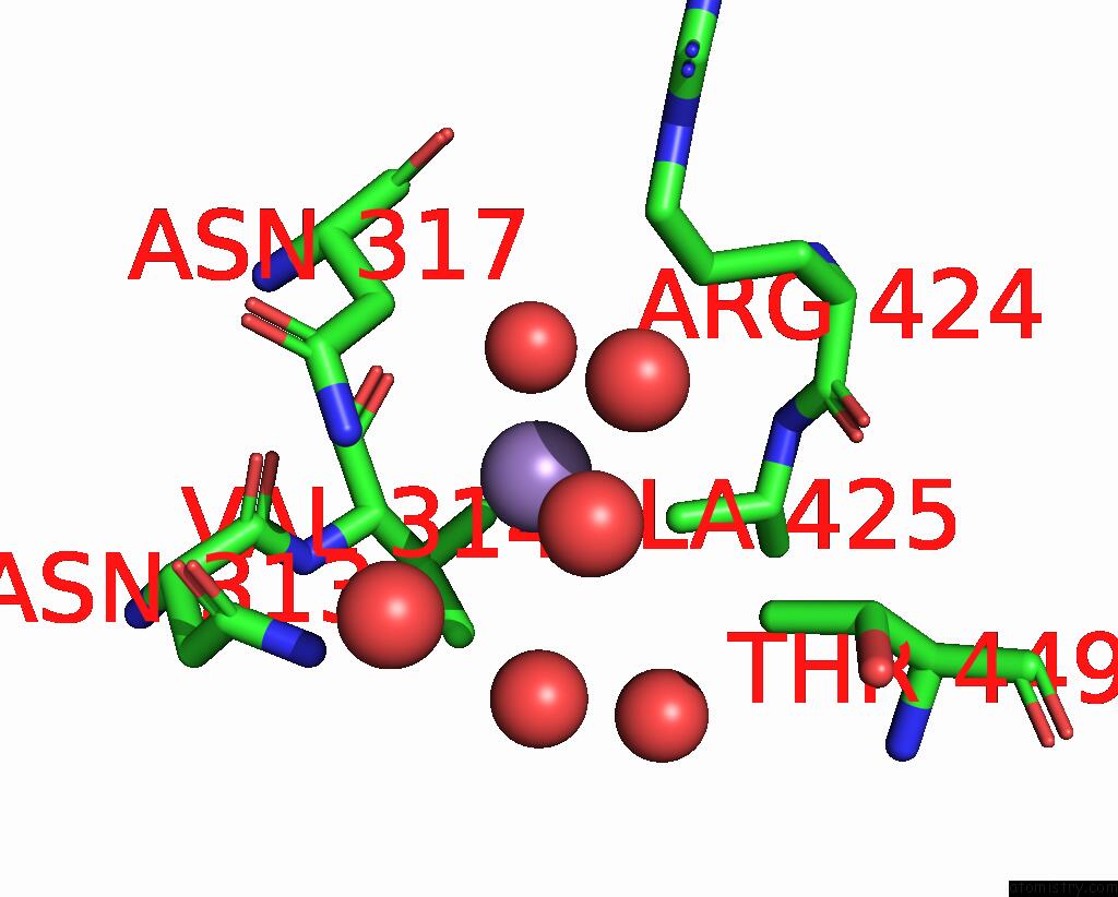
Mono view

Stereo pair view
Mono view
Stereo pair view
A full contact list of Manganese with other atoms in the Mn binding
site number 2 of Structural Analysis of PLD3 Reveals Insights Into the Mechanism of Lysosomal 5' Exonuclease-Mediated Nucleic Acid Degradation within 5.0Å range:
|
Reference:
Y.Roske,
C.Cappel,
N.Cremer,
P.Hoffmann,
T.Koudelka,
A.Tholey,
U.Heinemann,
O.Daumke,
M.Damme.
Structural Analysis of PLD3 Reveals Insights Into the Mechanism of Lysosomal 5' Exonuclease-Mediated Nucleic Acid Degradation. Nucleic Acids Res. 2023.
ISSN: ESSN 1362-4962
PubMed: 37994783
DOI: 10.1093/NAR/GKAD1114
Page generated: Sun Oct 6 13:38:46 2024
ISSN: ESSN 1362-4962
PubMed: 37994783
DOI: 10.1093/NAR/GKAD1114
Last articles
Zn in 9MJ5Zn in 9HNW
Zn in 9G0L
Zn in 9FNE
Zn in 9DZN
Zn in 9E0I
Zn in 9D32
Zn in 9DAK
Zn in 8ZXC
Zn in 8ZUF